 Live Science Plus


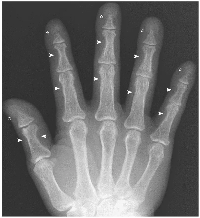
A man whose finger bones looked mysteriously "eaten away" on an X-ray was actually a textbook case for doctors who have seen what high levels of a hormone called parathyroid hormone can do to the body.
The 45-year-old patient in Japan had a noncancerous tumor on his parathyroid glands, which are four tiny glands that sit adjacent to the thyroid gland in the throat. The tumor was causing the glands to be overactive, and produce too much parathyroid hormone, which controls calcium levels in the body.
The man's high levels of the parathyroid hormone had led to an acceleration of the normal breakdown of his bones, resulting in shrinking finger bones. When doctors removed his tumor, the man's hormone levels quickly returned to normal, according to a report of his case published today (May 21) in the New England Journal of Medicine.
Article continues belowCases of overactive parathyroid glands that are as severe as this man's case are rarely seen nowadays. Instead, the condition is now usually caught before the hand bones begin to be affected to such an extent, experts say. [16 Oddest Medical Case Reports]
"This patient had a very high parathyroid hormone level, and a large tumor by today's standards, likely indicating long-standing and severe disease," said Dr. Bart Clarke, an endocrinologist at the Mayo Clinic in Rochester, Minnesota, who wasn't involved with the case.
It's rare for a tumor on the parathyroid glands to grow so large before getting diagnosed, Clarke said. "Occasional patients still show up with big tumors due to their choice to wait so long to have surgery, but these are unusual."
Hand X-rays used to be done routinely in patients suspected of having overactive parathyroid glands, also called hyperparathyroidism. But today, doctors diagnose the condition with blood tests. These tests have improved over the last 30 years, and eliminated the need for X-rays, Clarke told Live Science.
"Also, blood calcium level is checked more frequently than in the past, so milder forms of hyperparathyroidism are diagnosed more frequently in the U.S.," Clarke said.
The case illustrates how the bones in the body go through constant remodeling. One set of specialized cells break down bone and release calcium into the blood, which is called bone resorption. Meanwhile, other cells use the calcium to form new bone tissue.
"High levels of parathyroid hormone stimulate the bone resorbing cells to resorb bone more quickly, which leads to rapid transfer of more calcium into the blood," Clarke said. At the same time, the hormone signals to the kidneys to lower the calcium excreted from the body in the urine.
"Both processes lead to higher blood calcium," he said. "In this case, the high blood calcium can't go back into the bones because the increased parathyroid hormone level won't let it."
Other classical findings in patients with hyperparathyroidism include kidney stones, brown tumors in the bones and a condition called salt-and-pepper skull, in which areas of the skull bones become thin, and appear darker on X-rays.
Most of these findings often return to normal when overactive glands are removed, experts said.
Email Bahar Gholipour. Follow us @LiveScience, Facebook & Google+. Original article on Live Science.

