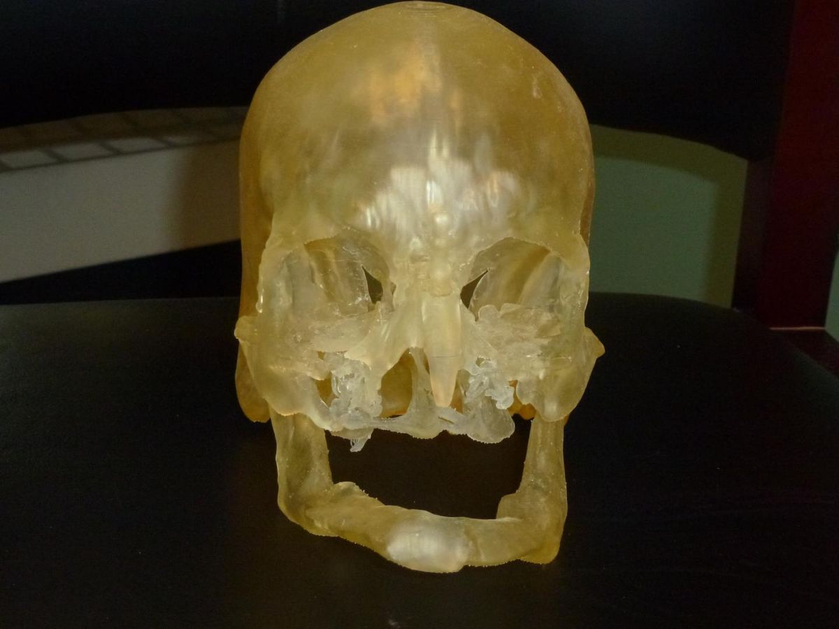
Surgeons are using new, highly accurate 3D printers to guide face transplantation operations, making the procedures faster and improving outcomes, according to a new report.
The face replicas made on these printers take into account bone grafts, metal plates and the underlying bone structure of the skull. They improve surgical planning, which ultimately makes the surgery much shorter, the report authors said.
The new technique has already been used in several patients, including two high-profile face transplant patients — Carmen Tarleton, who was maimed by her husband and received a face transplant in 2013, and Dallas Wiens, who was the first person in the U.S. to receive a full face transplant, in 2011.
The surgeries have dramatically improved the lives of the patients, the researchers said.
"They went from having no face and no features at all, to being able to talk and eat and breathe properly," said Dr. Frank Rybicki, a radiologist and the director of the Applied Imaging Science Laboratory at Brigham and Women's Hospital in Boston, who presented the findings today (Dec. 1) at the meeting of the Radiological Society of North America.
Custom fit
For the patients, face transplantation is often the end of a long journey.
Sign up for the Live Science daily newsletter now
Get the world’s most fascinating discoveries delivered straight to your inbox.
"Typically, by the time they come to us, they've had 20 or 30 surgeries already, just to save their lives," Rybicki told Live Science. [15 Odd Things That Can Be 3D Printed]
That means that patients may have plates, screws, bone grafts and dozens of other small modifications in their faces, and the new face has to fit perfectly around these. 3D printing allows the team to see exactly where these elements are, making the surgery — which can take up to 25 hours — go more quickly and smoothly, Rybicki said.
Soft tissue
The team printed out the soft tissue for Tarleton, whose estranged husband threw industrial-strength lye (a strong chemical used in soap making) on her face, according to the report.
The lye "literally burned off all the skin and all the squishy stuff in the face, and just left the bone," which was covered by a paper-thin flap of tissue, Rybicki said.
Printing soft tissue requires a sophisticated technique, but it was tremendously helpful because, without 3D printing, it's very difficult to visualize that tissue, Rybicki said.
Since her face transplantation procedure in 2011, Tarleton has done amazingly well, and her facial features have truly become her own, Rybicki said. The tissue has undergone dramatic remodeling, and the face no longer resembles neither her original face nor the donor's face. Now, three years after her operation, it is hard to tell that she was the recipient of a face transplant, Rybicki said.
Images of Tarleton's face will be revealed at the meeting later today.
The team also created 3D-printed versions of the new soft-tissue structure at Tarleton's follow-up appointments. As a result, they can document some of the facial remodeling that Tarleton has undergone, Rybicki said.
New innovations
Having a better understanding of the facial anatomy can also improve outcomes in less dramatic types of facial reconstruction, said Dr. Edward Caterson, a plastic surgeon at Brigham and Women's Hospital who is part of the same face transplant team.
For example, when someone's jaw is destroyed, doctors typically harvest a piece of rib or leg bone to replace the missing jaw. Because the tibia, or leg bone, is quite straight, it's tricky to cut it for a perfect fit. 3D printing allows that cut to be done more precisely, Caterson said.
"We're also getting an opportunity to innovate surgically, due to the fact we can do this planning preoperatively," Caterson told Live Science.
Recently, 3D printing enabled Caterson to harvest bone from a completely new location — the femur, or thigh bone. Though doctors often use rib grafts to replace jawbone, ribs don't have their own blood supply, so they typically collapse after a few years.
3D modeling allowed Caterson to use a portion of the femur that has its own blood supply, which should last much longer, he said.
Follow Tia Ghose on Twitter and Google+. Follow Live Science @livescience, Facebook & Google+. Originally published on Live Science.

Tia is the managing editor and was previously a senior writer for Live Science. Her work has appeared in Scientific American, Wired.com and other outlets. She holds a master's degree in bioengineering from the University of Washington, a graduate certificate in science writing from UC Santa Cruz and a bachelor's degree in mechanical engineering from the University of Texas at Austin. Tia was part of a team at the Milwaukee Journal Sentinel that published the Empty Cradles series on preterm births, which won multiple awards, including the 2012 Casey Medal for Meritorious Journalism.