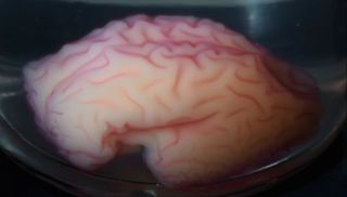
Scientists have discovered exactly how the human brain gets its crinkly, wrinkly appearance in utero.
It turns out that the huge explosion in the number of brain cells in the brain's outer layer, called the cortex, forces that layer to swell and then collapse in on itself to form those characteristic creases. This cortical origami — which has also evolved in a handful of other brainy species, such as dolphins and some primates — may be nature's way of solving the tight packing problem.
"This simple evolutionary innovation, with iterations and variations, allows for a large cortex to be packed into a small volume, and is likely the dominant cause behind brain folding, known as gyrification," said study co-author L. Mahadevan, an applied mathematician at Harvard University. [Video: See the Brain in a Vat Get Its Wrinkles]
Packed with brain cells
The outer surface of the human brain is a mass of curving bulges and fissures, known as gyri and sulci, all made of gray matter. Beneath this gray matter sits the white matter, the bundle of nerve fibers that send and receive signals between the brain and the rest of the body. Scientists have long suspected that this wrinkly brain surface evolved in order to squish many more brain cells, or neurons, into the relatively small space of the brain.
In the new study, the researchers decided to test their theory in a gel replica that more accurately reflected human brain anatomy. In the womb, the brain of a fetus starts out smooth, but between 14 weeks and 26 weeks of gestation, the fetal brain folds and curves in a predictable sequence from one brain region to the next, a 1997 study in the journal Ultrasound in Obstetrics and Gynecology found.
To recreate this process, the team collected magnetic resonance imaging (MRI) images of fetal brains and used those images to make an anatomically accurate gel reconstruction of the developing human brain.
Sign up for the Live Science daily newsletter now
Get the world’s most fascinating discoveries delivered straight to your inbox.
The team coated the outside layer of the mock brain with a stretchy elastomer gel to mimic the cortical layer. They placed this fetal-brain replica in a vat of solvent.
The brain quickly soaked up the solvent, and its outer layer ballooned outward more quickly than its inner layer. This uneven swelling caused compression and buckling, and within minutes, the team had recreated the gyri and sulci of the brain. What's more, the formation pattern was shockingly similar to that found in real brains, the researchers reported today (Feb. 1) in the journal Nature Physics.
"When I put the model into the solvent, I knew there should be folding, but I never expected that kind of close pattern compared to [the] human brain," said study co-author Jun Young Chung, a researcher in the Harvard University School of Engineering and Applied Sciences. "It looks like a real brain."
Interestingly, the team also showed that the size, shape and orientation of the largest folds were highly reliable, with the replica brain creasing in the same way no matter how many times it was put into the solvent. The smaller wrinkles appear to be unique, folding differently in each experiment. That mimics the pattern in human brains, in which the largest folding patterns are consistent across healthy people, but each individual has a unique pattern of little wrinkles.
Follow Tia Ghose on Twitterand Google+. Follow Live Science @livescience, Facebook & Google+. Original article on Live Science.

Tia is the managing editor and was previously a senior writer for Live Science. Her work has appeared in Scientific American, Wired.com and other outlets. She holds a master's degree in bioengineering from the University of Washington, a graduate certificate in science writing from UC Santa Cruz and a bachelor's degree in mechanical engineering from the University of Texas at Austin. Tia was part of a team at the Milwaukee Journal Sentinel that published the Empty Cradles series on preterm births, which won multiple awards, including the 2012 Casey Medal for Meritorious Journalism.