Tiny 'Ships' Sail Bloodstream to Destroy Cancer

Tiny ships loaded with a cargo that can identify and destroy cancerous tumors are being tested on mice and could one day ply the human bloodstream.
"These mother ships are only 50 nanometers in diameter, or 1,000 times smaller than the diameter of a human hair, and are equipped with an array of molecules on their surfaces that enable them to find and penetrate tumor cells in the body," explained biochemist Michael Sailor at the University of California, San Diego.
Sailor explains how these microscopic ships of destruction would sail through the body unnoticed: "The idea involves encapsulating imaging agents and drugs into a protective 'mother ship' that evades the natural processes that normally would remove these payloads if they were unprotected."
Metaphors and coincidental surnames aside, the idea is another important step in a broad effort by many researchers in several fields in recent years, all aimed at targeting tumors more efficiently by delivering no more drugs than necessary and getting them inside tumors so as not to destroy healthy cells.
"Many drugs look promising in the laboratory, but fail in humans because they do not reach the diseased tissue in time or at concentrations high enough to be effective," said co-researcher Sangeeta Bhatia, a physician and bioengineer at MIT. "These drugs don't have the capability to avoid the body's natural defenses or to discriminate their intended targets from healthy tissues. In addition, we lack the tools to detect diseases such as cancer at the earliest stages of development, when therapies can be most effective."
The hulls of the ships are created with specially modified lipids (a type of energy-storing fat) designed to mimic lipids that cover natural cells, allowing the ships to avoid the radar body's immune system. The stealth construction was tested on mice, and the ships sailed for hours before being destroyed.
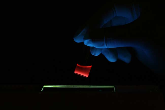
The ships were loaded with an anti-cancer drug plus superparamagnetic iron oxide and fluorescent quantum dots. The iron oxide nanoparticles allow the ships to show up in a Magnetic Resonance Imaging, or MRI, scan, while the quantum dots can be seen by a fluorescence scanner.
Sign up for the Live Science daily newsletter now
Get the world’s most fascinating discoveries delivered straight to your inbox.
"One can imagine a surgeon identifying the specific location of a tumor in the body before surgery with an MRI scan, then using fluorescence imaging to find and remove all parts of the tumor during the operation," Sailor said.
"This study provides the first example of a single nanomaterial used for simultaneous drug delivery and multimode imaging of diseased tissue in a live animal," said Ji-Ho Park, a graduate student in Sailor's laboratory who was part of the team.
The research, detailed in the journal Angewandte Chemie in Germany, was financed by a grant from the U.S. National Cancer Institute, which is part of the National Institutes of Health.
- The Top 10 Worst Hereditary Conditions
- Chemotherapy 'Bomb' Developed in Cancer War
- Video - Kids Health Issues
Robert is an independent health and science journalist and writer based in Phoenix, Arizona. He is a former editor-in-chief of Live Science with over 20 years of experience as a reporter and editor. He has worked on websites such as Space.com and Tom's Guide, and is a contributor on Medium, covering how we age and how to optimize the mind and body through time. He has a journalism degree from Humboldt State University in California.


