New View Inside Bacteria Could Improve Health
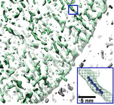
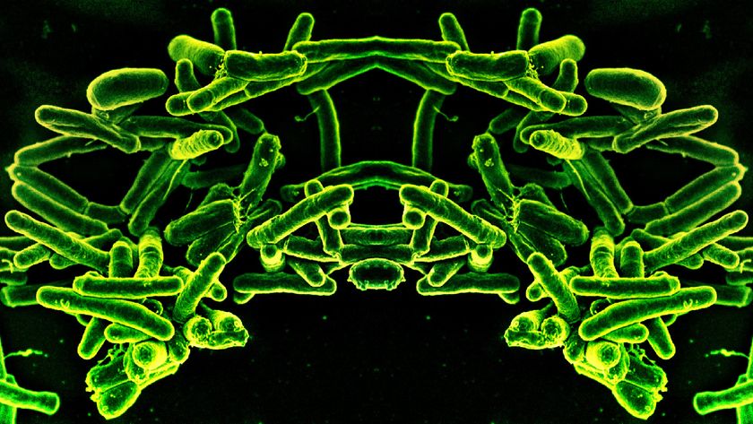
The bacterial cell wall, a primary target for the potent antibiotic penicillin, has been imaged for the first time in 3-D to show exactly how it gives bacteria their structural support and protection. Bacterial cells rely on a surrounding cage-like net, a molecular bag of sorts, to prevent rupturing and to maintain their structural strength, especially as they multiply. However, efforts to image such a tiny biological object was beyond technological reach until Caltech researchers received a gift from the Moore Foundation that allowed for the purchase of a new electron cryomicroscope that enabled them to be the first to visualize these biological structures at nanometer scales. "What we saw were long skinny tubes wrapping around the bag like the ribs of a person or a belt around the waist," said biologist Grant Jensen, the principal investigator on the study. "We also saw that the [bacterial cell wall] is just a single layer thick." That layer, called a sacculus, is made out of peptidoglycan, a mesh-like structure of carbohydrates (glycans) and amino-acid peptides. It is the sacculus, Jensen notes, that is targeted by the antibiotic penicillin (animal cells do not have cell walls); penicillin blocks a bacterium's ability to remodel its surrounding molecular bag as the bacterium itself grows. "If the bug can't make this bag," Jensen said, "it can't multiply, and you get better." Now that scientists can see how a bacterial cell wall is physically constructed, said Jensen, they’re closer to understanding "how a bacterium could direct its own growth, and how drugs that block that process might work." The research is detailed in the online early edition of the journal Proceedings of the National Academy of Sciences (PNAS).
- Gallery: Microscopic Images As Art
- Top 10 Mysterious Diseases
- New Approach Disarms Deadly Bacteria
Sign up for the Live Science daily newsletter now
Get the world’s most fascinating discoveries delivered straight to your inbox.

