Art Meets Science in Amazing Images
Plant Seed
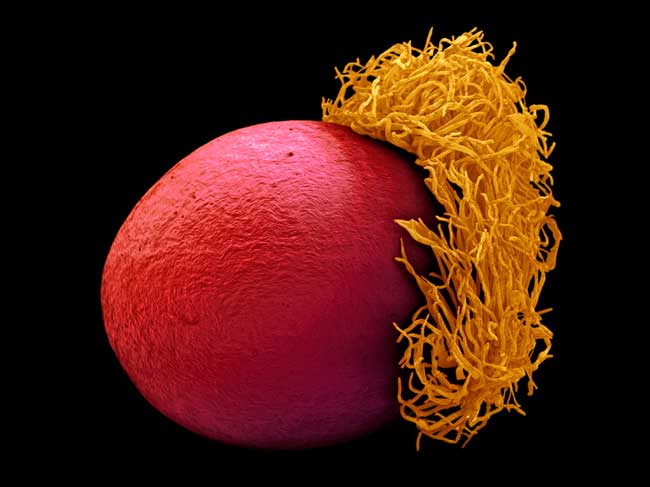
This seed is from a bird of paradise plant (Strelitzia reginae). Native to South Africa, the plant has a distinctive orange and blue flower, resembling an exotic bird, from which it takes its name. This image received an award from the Wellcome Trust, as part of the annual Wellcome Image Awards, for its ability to communicate the wonder and fascination of science.
Skin Cell
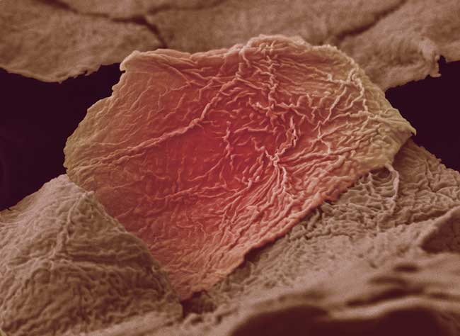
This image shows damaged skin cells from a hand (Anne Weston's own) scalded by boiling soup. The original image was black and white; the pink color has been added later. This image received an award from the Wellcome Trust, as part of the annual Wellcome Image Awards, for its ability to communicate the wonder and fascination of science.
Engineering DNA
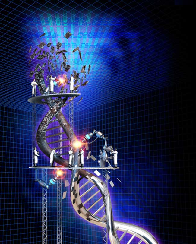
A strand of DNA, is modeled and rendered in 3D software to show how DNA is being modified and corrected. The rough, rusty metal is at the top and it's being chrome-plated as you go down and manipulated. This image received an award from the Wellcome Trust, as part of the annual Wellcome Image Awards, for its ability to communicate the wonder and fascination of science.
Sickle Cell
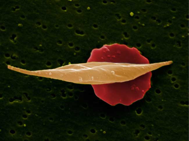
This image shows two red blood cells. The one in the front has been affected by sickle-cell anemia, and displays the characteristic sickle shape (a flattened C shape) common to the disease. This image received an award from the Wellcome Trust, as part of the annual Wellcome Image Awards, for its ability to communicate the wonder and fascination of science.
Summer Plankton
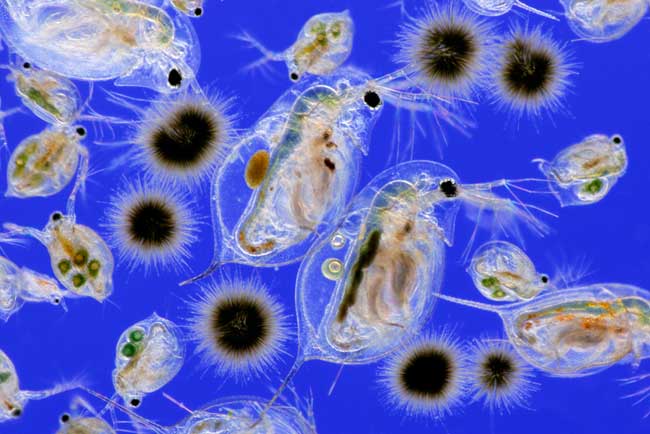
This photograph shows sea organisms known plankton, small organisms that drift in the oceans, seas and fresh water. Many types of plankton, such as these, are microscopic, but some, such as jellyfish, are very large. This image received an award from the Wellcome Trust, as part of the annual Wellcome Image Awards, for its ability to communicate the wonder and fascination of science.
Compact Bone
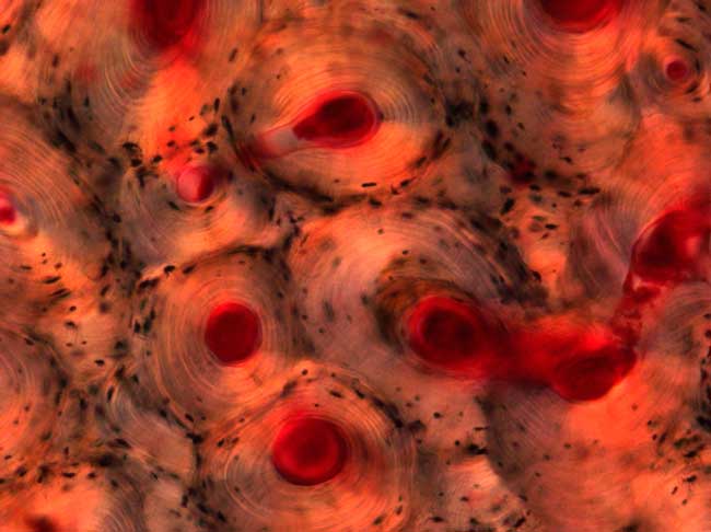
These circular structures are regions of compact bone from a human femur. Compact bone forms a hard outer shell around the spongy bone that makes up the marrow space in the center. This image received an award from the Wellcome Trust, as part of the annual Wellcome Image Awards, for its ability to communicate the wonder and fascination of science.
Cancer Cell
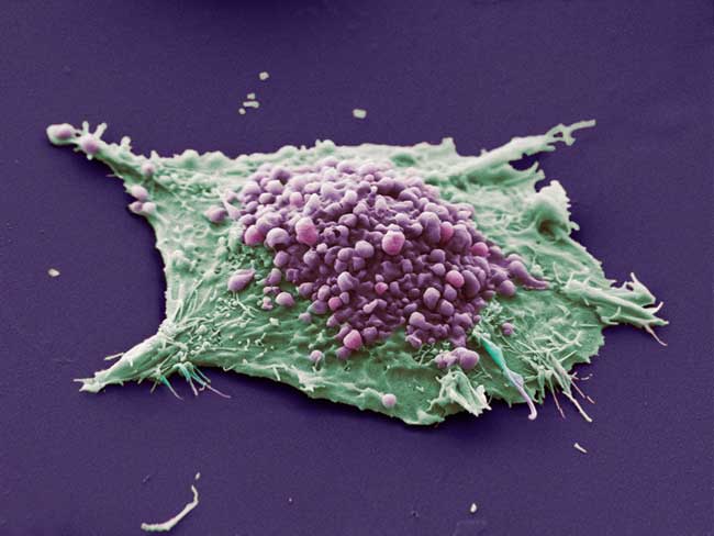
This image shows a single cell grown from a culture of lung epithelial cancer cells. The purple spheres are blebs: irregular bulges where the cell's internal scaffolding - its cytoskeleton - becomes unlinked from the surface membrane. This image received an award from the Wellcome Trust, as part of the annual Wellcome Image Awards, for its ability to communicate the wonder and fascination of science.
Get the world’s most fascinating discoveries delivered straight to your inbox.
Tibetan Doctor

Amchi Tala, a Tibetan doctor, holds precious medical books in a remote region of Western Tibet. The items include the Gyu Shi (Four Tantras), the fundamental Tibetan medical classic written in the 12th century, and a manuscript on compounding medicines, written by previous generations of Amchi Tala's medical family. This image received an award from the Wellcome Trust, as part of the annual Wellcome Image Awards, for its ability to communicate the wonder and fascination of science.
Vitro Egg
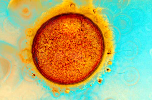
This image shows sperm and an egg (or ovum) at the moment of conception by in vitro fertilization (IVF). The egg is surrounded by protective cumulus cells around the outside surface, colored yellow. The sperm need to penetrate the membrane surrounding the egg, called the zona pellucida, if successful fertilization is to occur. This image received an award from the Wellcome Trust, as part of the annual Wellcome Image Awards, for its ability to communicate the wonder and fascination of science.
Mouse Head
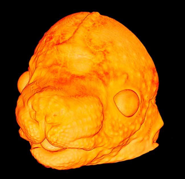
This 3D image depicts a developing embryonic mouse head at age 14.5 days. The pores of future whiskers are visualized as small bumps across the snout and various vessels connecting the eye, including the optic nerve, are clearly observed, but it is clear at this stage that the eyelids have yet to form. This image received an award from the Wellcome Trust, as part of the annual Wellcome Image Awards, for its ability to communicate the wonder and fascination of science.
Mouse Liver
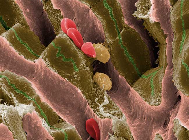
This image of the liver shows blood vessels called sinusoids as long pink channels, brown tissue that is important in the production of bile. The channels - shown as thin green grooves - carry the bile towards the small intestine to help digestion. This image received an award from the Wellcome Trust, as part of the annual Wellcome Image Awards, for its ability to communicate the wonder and fascination of science.


