1st complete map of an insect's brain contains 3,016 neurons
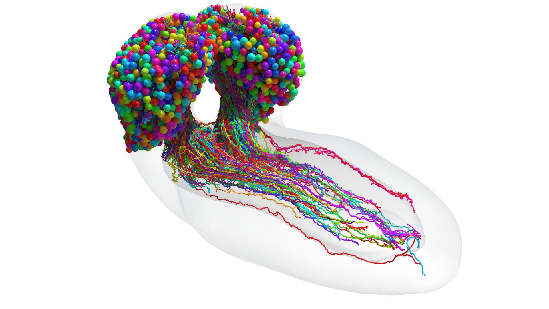
Scientists created a map of an entire larval fruit fly brain that shows all 548,000 synapses in the organ.

Scientists have unveiled the first complete map of an insect's brain.
The comprehensive map, called a connectome, took 12 years of meticulous work to construct, and shows the location of all 3,016 neurons in the brain of a larval fruit fly (Drosophila melanogaster). Between those brain cells are 548,000 points of connection, or synapses, where cells can send each other chemical messages that, in turn, trigger electrical signals that travel through the cells' wiring.
Researchers identified networks through which neurons on one side of the brain send data to the other, the team reported March 9 in the journal Science. The team also classified 93 distinct types of neurons, which differ in their shape, proposed function and the way they connect to other neurons.
The new connectome is remarkable for its completeness, experts told Live Science.
Related: Google just created the most detailed image of a brain yet
"This study is the first to be able to map the entirety of an insect central brain and thus characterize all synaptic pathways of all the neurons," Nuno Maçarico da Costa and Casey Schneider-Mizell, members of the Neural Coding group at the Seattle-based Allen Institute for Brain Science who were not involved in the initiative, told Live Science in a joint email.
In 2020, a different research group published a partial connectome of an adult fruit fly that contained 25,000 neurons and 20 million synapses. But scientists have complete connectomes for only three other organisms: a nematode, a larval sea squirt and a larval marine worm. Each of those connectomes contains a few hundred neurons and lacks the distinct brain hemispheres seen in insects and mammals, said study co-senior author Joshua Vogelstein, director and co-founder of the NeuroData lab at Johns Hopkins University.
Sign up for the Live Science daily newsletter now
Get the world’s most fascinating discoveries delivered straight to your inbox.
More than 80 people helped to construct the new connectome, study first author Michael Winding, a research associate in the University of Cambridge Department of Zoology, told Live Science in an email. To do so, scientists thinly sliced a larval fly brain into 5,000 sections and snapped microscopic images of each slice. They pieced these images together to form a 3D volume. The team then pored over the images, identified individual cells within them and manually traced their wires.
The resulting map surprised scientists in several ways.
For example, scientists tend to think of neurons sending outgoing messages through long wires called axons and receiving messages through shorter, branched wires called dendrites. However, there are exceptions to this rule, and it turns out that axon-to-axon, dendrite-to-dendrite and dendrite-to-axon connections make up about one-third of the synapses in the larval fly brain, Winding said.
Related: How does the brain store memories?
The connectome was also surprisingly "shallow," meaning incoming sensory information passes through very few neurons before getting passed to one involved in motor control, which can direct the fly to perform a physical behavior, Vogelstein said. To achieve this level of efficiency, the brain has built-in "shortcuts" between circuits that somewhat resemble those in state-of-the-art AI systems, Winding said.
One limitation of the connectome is that it doesn't capture which neurons are excitatory, meaning they push other neurons to fire, or inhibitory, meaning they make neurons less likely to fire, Schneider-Mizell said. These dynamics affect how information flows through the brain, he said.
Still, the connectome opens the door to many future advancements, such as more energy-efficient AI systems and a better understanding of how humans learn, Vogelstein said.
"Humans do things such as make decisions, learn, navigate the environment, eat," he said. "And so do flies. And there's good reason to think that the mechanisms that flies have for implementing those kinds of cognitive functions are also in humans."

Nicoletta Lanese is the health channel editor at Live Science and was previously a news editor and staff writer at the site. She holds a graduate certificate in science communication from UC Santa Cruz and degrees in neuroscience and dance from the University of Florida. Her work has appeared in The Scientist, Science News, the Mercury News, Mongabay and Stanford Medicine Magazine, among other outlets. Based in NYC, she also remains heavily involved in dance and performs in local choreographers' work.


