Primitive Eye, Tiny Liver Grown in the Lab
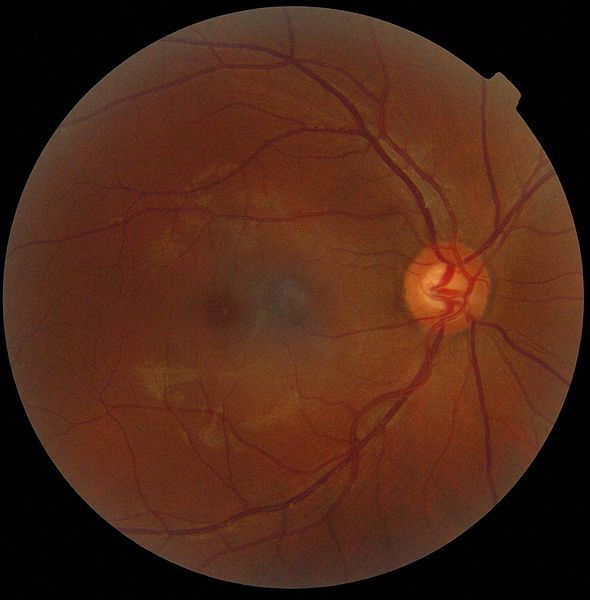
Japanese scientists claim to have coaxed stem cells to develop into a rudimentary human liver, replete with working blood vessels and the ability to metabolize. At the same time, another group in Japan reports the growth from stem cells of a precursor of a human eyeball.
Both feats were presented at the annual meeting of the International Society for Stem Cell Research in Yokohama, Japan, last week. Although further progress is needed before fully functional lab-grown livers and eyes will be ready to implant into a human, outside experts say the new results constitute genuine advances in that direction — and they have other medical uses in the meantime.
Takanori Takebe, a stem-cell biologist at Yokohama City University in Japan, and his team grew a small, rudimentary liver using a recipe of just three types of cells. The trick was figuring out when to introduce each ingredient into the mix of cells: "It took over a year and hundreds of trials," Takebe was quoted as saying in Nature.
First, the researchers placed genetically reprogrammed human skin cells, called "induced pluripotent stem cells," on growth plates in a specially designed chemical bath. After nine days, the cells began developing into hepatocytes, or liver cells. At that point, the researchers added cells taken from an umbilical cord, which would develop into the lining of blood vessels, and cells from bone marrow that can differentiate into bone, cartilage or fat.
Two days later, the cell assortment had self-organized to form a three-dimensional "liver bud" — a 5-millimeter-wide chunk of tissue that performed basic liver functions. When they grafted the liver bud into a mouse, the researchers said the tiny organ's blood vessels worked correctly, and it successfully metabolized some drugs that human livers metabolize but which mouse livers normally cannot. [How to Build a Human Brain]
Takebe said a more developed version of the liver could eventually be used for long-term organ replacement, as well as serving as a short-term graft for patients whose damaged native livers are expected to recover. Much more work is needed to get to that point, however, as the liver bud lacks critical features called bile ducts, and its cells do not produce as much of a plasma protein called albumin as natural liver cells. Still, the group's efforts thus far "blew my mind," said George Daley, director of the stem-cell transplantation program at the Boston Children's Hospital in Massachusetts.
Meanwhile, Yoshiki Sasai and colleagues at the RIKEN Center for Developmental Biology in Kobe, Japan, reported that they had managed to induce human stem cells called "retinal precursor cells" to develop into a central component of the human eye called an optic cup. In a petri dish, the cells spontaneouslybulged to form a bubble called an eye vesicle, which folded back on itself to create a half-millimeter-wide pouch layered with retinal cells — the optic cup.
Sign up for the Live Science daily newsletter now
Get the world’s most fascinating discoveries delivered straight to your inbox.
Most impressively, according to outside researchers, this process unfolded correctly without any external guidance by the researchers. "The morphology is the truly extraordinary thing," said Austin Smith, director of the Centre for Stem Cell Research at the University of Cambridge in the U.K. In fact, scientists didn't even know quite how optic cups developed until Sasai and his team watched it happen spontaneously in the lab.
Researchers say the achievement lends hope to the possibility of restoring vision in humans who have lost their sight. Masayo Takahashi, an ophthalmologist on Sasai's team, has already started transferring sheets of retina from lab-grown optic cups into blind mice in hopes of restoring their vision, and she plans to do the same with monkeys by the end of the year. [The 6 Craziest Animal Experiments]
The open question is whether transplanted tissue will integrate into native tissue. Sasai insists that the group's optic cups are "pure," meaning they do not contain any leftover embryonic stem cells, which pose a risk of developing into cancerous growths or unrelated tissues. "You'd have no more reason to expect bone to be growing in these eyes," he said.
Stem cells have successfully been used to generate functional human windpipes and bladder walls, and several animal organs have been grown from stem cells in the lab, including lungs and penises.
Follow Natalie Wolchover on Twitter @nattyover. Follow Life's Little Mysteries on Twitter @llmysteries. We're also on Facebook & Google+.
Natalie Wolchover was a staff writer for Live Science from 2010 to 2012 and is currently a senior physics writer and editor for Quanta Magazine. She holds a bachelor's degree in physics from Tufts University and has studied physics at the University of California, Berkeley. Along with the staff of Quanta, Wolchover won the 2022 Pulitzer Prize for explanatory writing for her work on the building of the James Webb Space Telescope. Her work has also appeared in the The Best American Science and Nature Writing and The Best Writing on Mathematics, Nature, The New Yorker and Popular Science. She was the 2016 winner of the Evert Clark/Seth Payne Award, an annual prize for young science journalists, as well as the winner of the 2017 Science Communication Award for the American Institute of Physics.

