Beauty and Brains: Award-Winning Medical Images
Leaf of Lavender
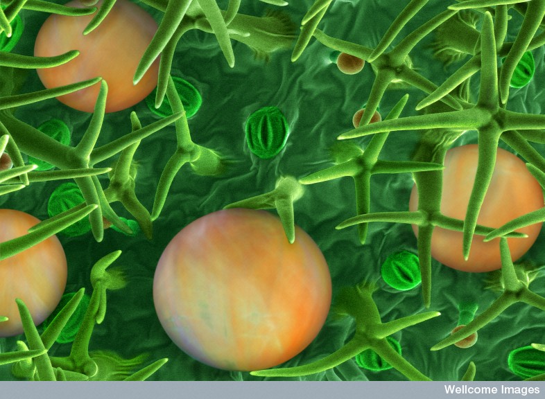
This false-coloured scanning electron micrograph (SEM) shows a lavender leaf (Lavandula) imaged at 200 microns. The surface of the leaf is densely covered with fine hair-like outgrowths made from specialised epidermal cells called non-glandular trichomes.
Frog Oocytes
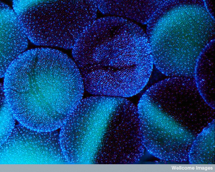
This confocal micrograph shows stage V-VI oocytes (800-1000 micron diameter) of an African clawed frog (Xenopus laevis), a model organism used in cell and developmental biology research. Each oocyte is surrounded by thousands of follicle cells, shown in the image by staining DNA blue. Blood vessels, which provide oxygen to the oocyte and follicle cells, are shown in red. The ovary of each adult female Xenopus laevis contains up to 20 000 oocytes. Mature oocytes are approximately 1.2 mm in diameter, much larger than the eggs of many other species.
A Cancer Cell Divides
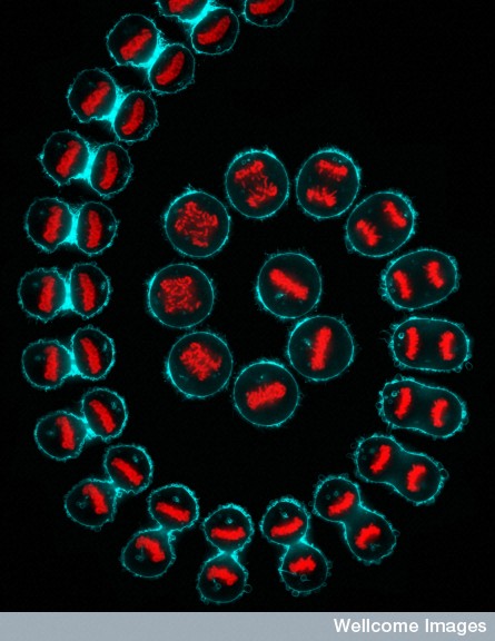
This composite confocal micrograph uses time-lapse microscopy to show a cancer cell (a HeLa cell derived from the cancer of a woman named Henrietta Lacks) undergoing cell division (mitosis). The DNA is shown in red, and the cell membrane is shown in cyan.
Stunning Seedling
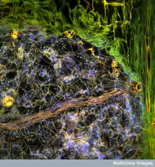
This confocal micrograph shows the tissue structures within the leaf of an Arabidopsis thaliana seedling. The sample was fixed and stained with propidium iodide, which labels DNA, but was imaged four years later. Different oxidation of the staining chemical in different tissues allows researchers to investigate the structures within.
Caffeine Crystal
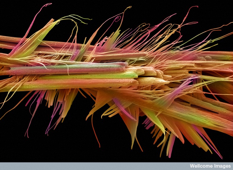
This false-coloured scanning electron micrograph shows caffeine crystals. Caffeine is found occurring naturally in plants, where its bitterness serves as a defense mechanism.
Chicken Embryo
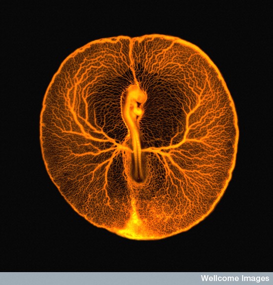
This fluorescence micrograph shows the vascular system of a developing chicken embryo (Gallus gallus), two days after fertilization.
Moving Cancer Cells
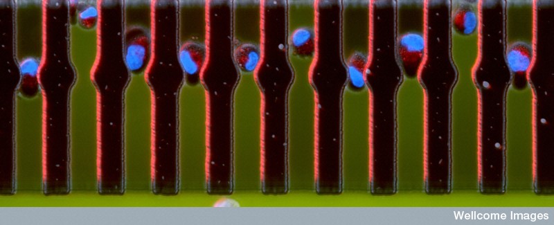
Taken in the course of research into how cancer cells move and spread, this Wellcome honoree shows cancer cells traveling through spaces a tenth the width of a human hair.
Sign up for the Live Science daily newsletter now
Get the world’s most fascinating discoveries delivered straight to your inbox.
Moth Fly
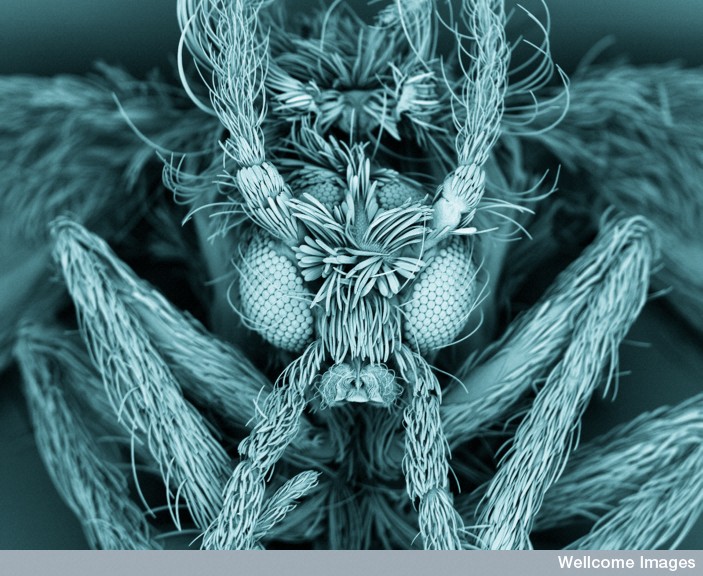
This false-colored image of a moth fly reveals the insect's fuzzy body and compound eyes.
Diatom Case
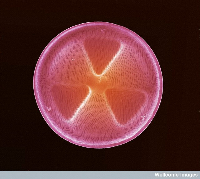
This false-colored scanning electron micrograph shows a diatom frustule. Diatoms are unicellular organisms and a major group of algae. Diatoms are encased within a hard cell wall made from silica. Frustules have a variety of patterns, pores, spines and ridges, which are used to determine genera and species. Diatoms are one of the most common types of phytoplankton, and their communities are often used to measure environmental conditions such as water quality. This diatom is approximately 80 microns in diameter.
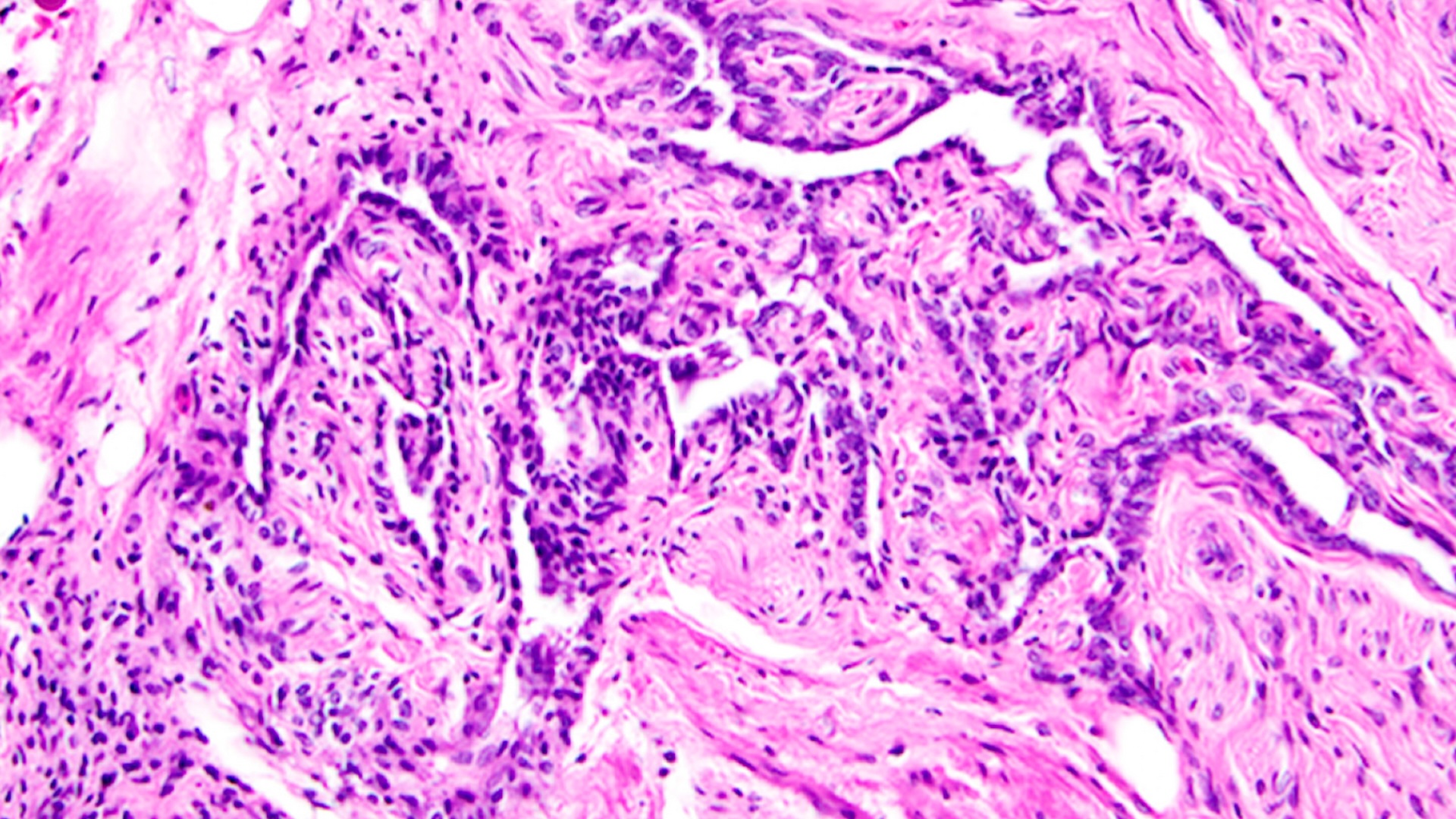
Hole in the Heart
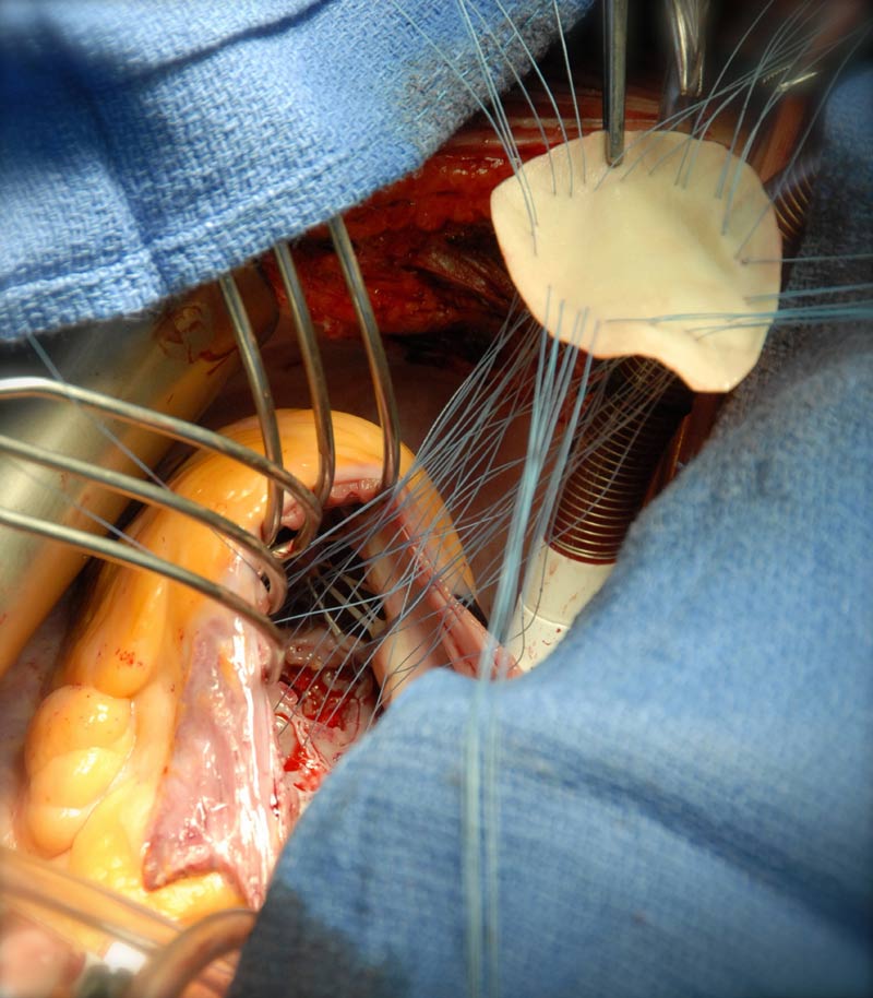
This photograph shows the repair of a traumatic ventricular septal defect (VSD). A VSD is a hole between the right and left ventricles of the heart, and is usually seen as a congenital condition, known as a 'hole in the heart'. This picture was taken in theatre to document the unusual injury and its subsequent repair; the VSD is seen at the bottom of this image, and a bovine patch is being stitched and parachuted into place to seal the defect.
Bacteria Biofilm
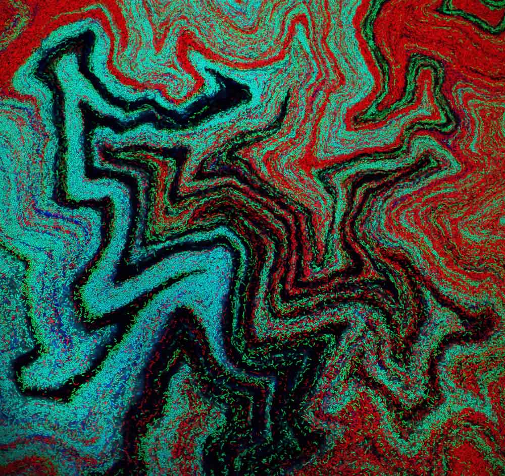
This micrograph photo was taken as part of a synthetic biology project and shows Bacillus subtilis, a Gram-positive, rod-shaped bacterium that is commonly found in soil. Distinct lineages of bacteria expressing different fluorescent proteins were initially mixed randomly on a petri dish. As the bacteria grow, they organize themselves into reproducible patterns and shapes that can be predicted with mathematical models.


