Award-Winning Microscope Images
Top prize
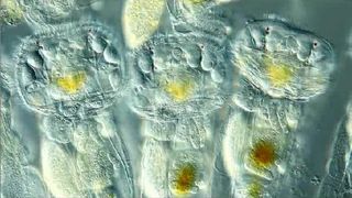
The top prize in the 2012 Olympus BioScapes competition went to Ralph Grimm, a teacher from the Australian town of Jimboomba, just south of Brisbane, for his 58-second video of tiny animals called rotifers on a lily leaf in his pond. This still from Grimm's footage shows the creatures' trembling cilia, internal organs and eyes, which look like red dots.
Red alga
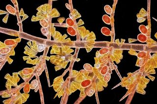
Arlene Wechezak of Anacortes, Wash., took second place with her up-close image of branching red alga, showing the tiny plant's reproductive tetraspores and golden diatoms.
Spectacular spore factory

Third prize went to Igor Siwanowicz, of the Howard Hughes Medical Institute's Janelia Farm research campus in Virginia, for his colorful shot of spore-filled sporangia on a fern (Polypodium virginianum).
Tiny crustacean claw
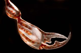
This image of a translucent claw of a pram bug, with muscles and rows of pigment cells visible, earned fourth prize.
Single-cell wonder

This picture is actually made up of 22 stacked images of the unicellular green alga Micrasterias taken from a lake sample. It came in 5th place.
Coral mouth
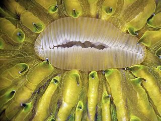
This 6th prize image offers a glimpse into the mouth of a live mushroom coral (Fungia sp.) as it expands.
Fly brain

The image shows the brain of a fruit fly larva with eye discs attached. It took 7th place.
Sign up for the Live Science daily newsletter now
Get the world’s most fascinating discoveries delivered straight to your inbox.
Up-close deadnettle
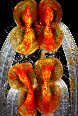
The 8th-prize shot shows the stamens anthers and filaments on a type of deadnettle.
Flower seed
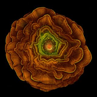
Striking in itself, this Delphinium seed gives rise to beautiful flowers. This image nabbed the 9th prize.
Butterfly scales

The wing scales of a Prola Beauty butterfly are showcased in this microscopic 10th-place image.

Most Popular


