Making Maps In The Brain
(ISNS) -- Disorientation is often one of the first warnings of Alzheimer's disease. Someone gets into the car to go to the grocery store, and suddenly can't remember how to get there. Now, neurologists offer a clue as to why the first kind of memory to fade may be navigational.
Researchers at Emory University near Atlanta have demonstrated that primates map their environment using "grid cells," specialized neurons that help the animals navigate by overlaying a virtual grid, made of triangles, atop regularly spaced points in the environment.
Elizabeth Buffalo, the study's lead author, is an associate professor of neurology at Emory University School of Medicine. She suspects that these grid cells don't just help primates navigate -- they also help form visual memories. And because of their location in the brain, they are especially susceptible to the ravages of Alzheimer's.
Grid cells were identified for the first time in 2005, by a group of scientists in Norway interested in how the brain enables animals to navigate. They put six rats in a pen, and monitored individual neurons while the rats explored.
The neurons they were watching lie in a part of the brain called the entorhinal cortex. It sits in the lower part of the brain, near its intersection with the brain stem. This is the perfect position for a mapmaker: the entorhinal cortex gets input about the environment from the senses, and sends its output up to the hippocampus, which is known for its roles in memory and navigation.
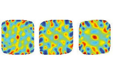
As a rat walked around the enclosure, a neuron in the entorhinal cortex fired; the rat walked a little more, and the neuron fired again. When the team mapped out all the points in the enclosure that had lit up a particular neuron, they found that these weren't just random signals: those "hot spots" defined a grid of equilateral triangles laid side by side.
The grid produced by each grid cell can serve as a basic map, where the hot spots are like signposts. By arranging these signposts in triangles, the brain can fit in more of them than it could if they were the same distance apart, but arranged in rectangles.
Sign up for the Live Science daily newsletter now
Get the world’s most fascinating discoveries delivered straight to your inbox.
Grid cells are distributed irregularly in the entorhinal cortex, and each one produces a slightly different grid. These grids overlap to generate a high-resolution map of the entire environment.
In humans, the entorhinal cortex is one of the first areas to degenerate in Alzheimer's disease. While experiments using functional magnetic resonance imaging had hinted at the presence of human grid cells, they had never been directly observed in any primate.
Buffalo's experiment changed that. In research reported in November in the journal Nature, three Rhesus monkeys looked at images on a computer screen while tiny microelectrodes monitored neurons in the entorhinal cortex.
When Buffalo and her coworkers compared eye-tracking results to the electrode measurements, they found that the monkeys, like the rats, were using neurons in the entorhinal cortex to construct a triangular grid they could superimpose on their environment.
Primates, though, are more sophisticated cartographers: the monkeys were able to activate their grid cells simply by looking around.
"We tend to explore things with our eyes," said Buffalo, and unlike the rats in the original experiments, "primates don't have to actually visit a place to construct the same kind of mental map."
Showing the monkeys the same picture twice enabled Buffalo to link grid cells to memory. When the monkeys looked at a familiar image, some cells fired less frequently, apparently remembering what they had already mapped. This suggests that grid cells may provide "a kind of framework for making associations," said Buffalo. The grid becomes the scaffold on which animals construct their visual memories.
That has important implications.
One of Buffalo's research interests is the early diagnosis of neurodegenerative diseases. Studies of brain changes in Alzheimer's disease in humans consistently show localized degeneration in the same parts of the entorhinal cortex where Buffalo found grid cells in monkeys.
May-Britt Moser, one of the authors of the original Norwegian study, described Buffalo's results as "extremely exciting." She suspects that the cells Buffalo observed, which respond to the monkeys' eye movements, may represent a new type of grid cell -- and that grid cells may start turning up in a variety of neurological contexts.
In the brain,"what works will be used over and over again," said Moser.
The next step is to study grid cells in a 3-D virtual environment, where the ability to manipulate the monkeys' surroundings permits researchers to study how grid cells respond to a range of variables.
"Now that we've identified them, there are so many questions we can ask," Buffalo said.
Eleanor Nelsen is a science writer based in Madison, Wisconsin.
Inside Science News Service is supported by the American Institute of Physics.
