Inside a Gross Anatomy Lab: The Human Body as Textbook
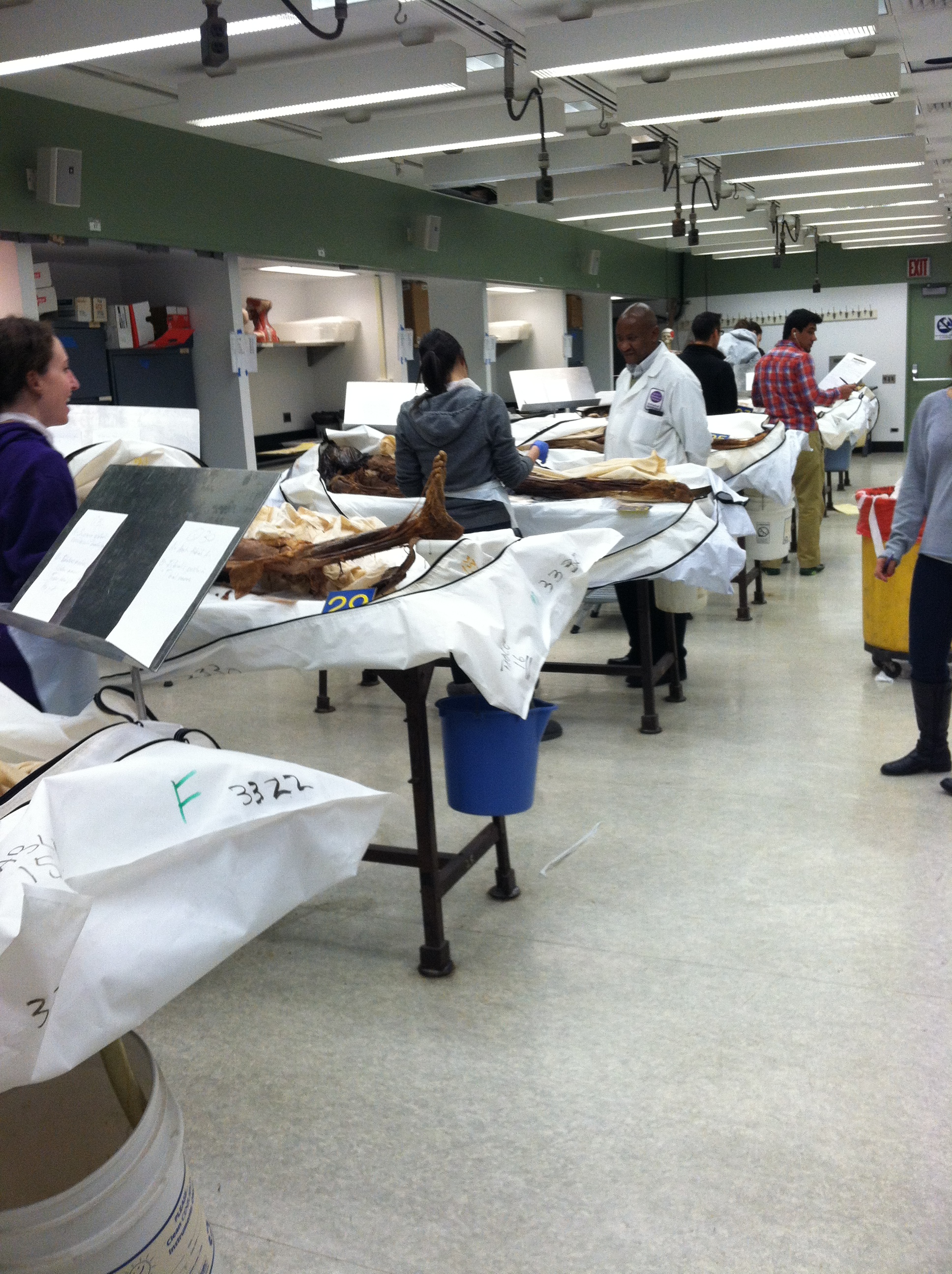
NEW YORK — The first things my eyes land on are the leg bones. Thin, sinewy strips of muscle and skin cling delicately to the femurs, tibias and fibulas. The feet have more flesh on them. And toenails.
About a dozen partially draped human cadavers lie atop dissecting tables in the basement gross anatomy lab here at NYU School of Medicine. Each of these individuals — mothers, fathers, grandparents, siblings — made the most admirable of gifts: donating their bodies to science.
I've been mentally preparing for this moment since I made the decision several weeks ago to visit a gross anatomy lab, to get a glimpse of a rite of passage medical students have undergone for hundreds of years. For many, it is their first experience with dead bodies, and the donor is their first patient. Yes, the students will learn the names and locations of all the major bones, muscles and organs in the body ― but they will also learn things a textbook could never teach them: the variability among human bodies, and the emotional connection that comes with being a doctor.
Seeing the donors' bodies doesn't have the disturbing effect on me I expected. I've watched surgeries before, and those times, the sight of exposed human flesh hit me on a visceral level — making me feel lightheaded and faint. Yet now, as the bodies lie here so peacefully and so clearly uninhabited, I get a strange feeling of calm and detachment. [The 10 Weirdest Ways We Deal With the Dead]
The smell hits me next. The chemicals used to preserve the bodies give off an odor that's somewhere between shoe polish and a mossy, earthen smell. Not exactly pleasant, but not noxious, either ― just ever-present, imprinting itself on my memory. It's not just formaldehyde. "Each medical school has its own special brew," lab instructor Melvin Rosenfeld, the university's associate dean for medical education, tells me.
Today's lab is not for medical students, but for physician assistants (PAs) from Pace University. To my relief, the PA students aren't required to do the dissecting themselves. Instead, the bodies have been prepared for them ahead of time and tagged with cards bearing instructions like, "Identify this muscle."
I approach one of the students, a young woman named Dominique Sisto, as she works. What does she think about working with the donors? "I'm grateful to them," Sisto says. "It lets you get up close and personal with the human body."
Sign up for the Live Science daily newsletter now
Get the world’s most fascinating discoveries delivered straight to your inbox.
Finally, I decide it's time to get up close and personal myself. Rosenfeld leads me, gloves donned, over to one of the donors. She's female and fairly petite, and her head remains covered. "Do you want to see the organs?" Rosenfeld asks, already reaching down and withdrawing one of the donor's lungs. He lets me hold it. It's much firmer and denser than I imagined a lung to be, though that's partly because of the fixative, which stiffens and preserves the tissue. I try to imagine it filling and compressing inside a living person.
Next, Rosenfeld picks up the heart. They tell you the heart is a muscle, and looking at this one, it's undeniable. All of a sudden, I'm cupping the heart in my hand. I can't believe this organ, about the weight and shape of a mango, once powered a human being. Rosenfeld excitedly points out some flimsy-looking fibers known as the tendinous cords (chordae tendineae) — literally, the heart strings — and explains how they are actually very strong and prevent the backflow of blood through the heart valves.
In that moment, as I'm standing there holding the heart, Rosenfeld is just a teacher, and I am just a student, and this body in front of us is the world's most beautiful textbook.
Follow Tanya Lewis on Twitter and Google+. Follow us @livescience, Facebook & Google+. Original article on LiveScience.com.
