Au. Sediba Gallery: Anatomy of Humanity's Closest Relative
Sediba's Skull
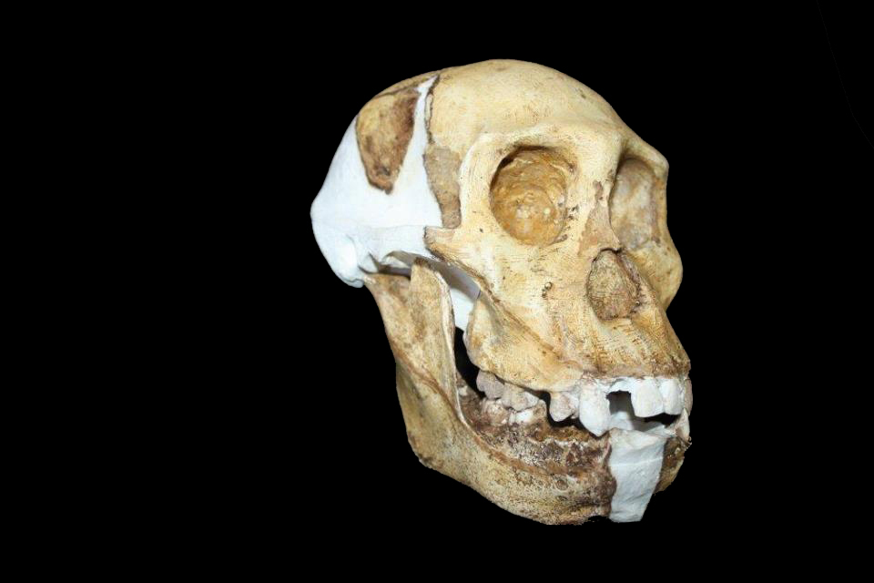
A team of scientists have completed the most detailed investigation of the anatomy of what may be the immediate ancestor of the human lineage, called Australopithecus sediba, shedding light on secrets about how it might have behaved.
The reconstructed skull and mandible of Au. sediba.
Au. sediba composite
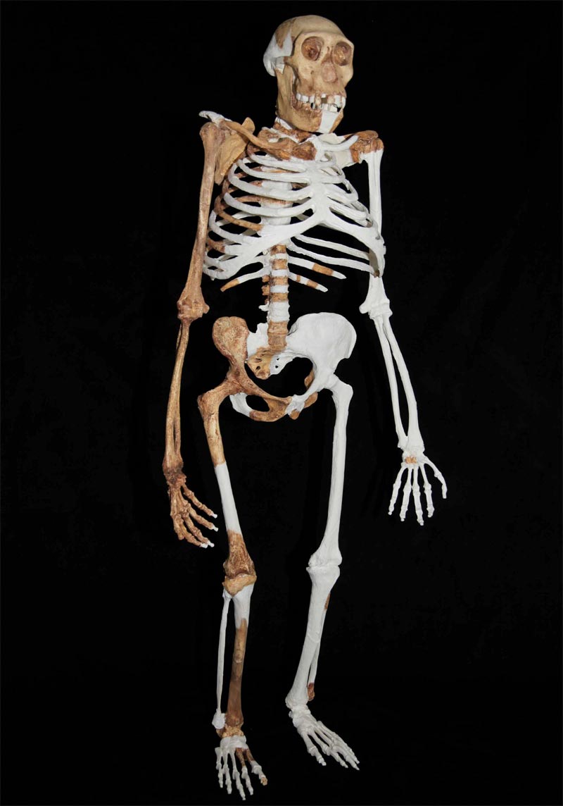
Composite reconstruction of Au. sediba based on recovered material from a younger male skeleton (MH1), a female skeleton (MH2) and an adult (MH4), and based upon the research presented in the accompanying manuscripts in the journal Science. As all individuals recovered to date are approximately the same size, size correction was not necessary. Femoral length was established by digitally measuring a complete femur of MH1 still encased in rock.
Skeletal comparison
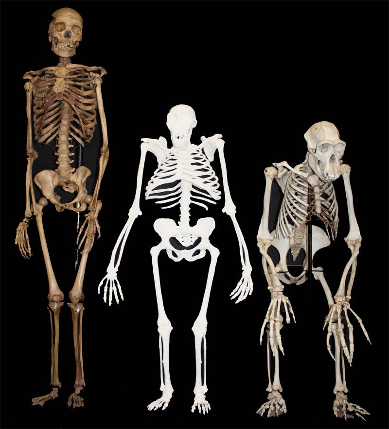
Another composite reconstruction of Au. sediba, with a small-bodied female modern H. sapiens skeleton (left) and a male Pan troglodytes (right) for comparison.
Upper limbs
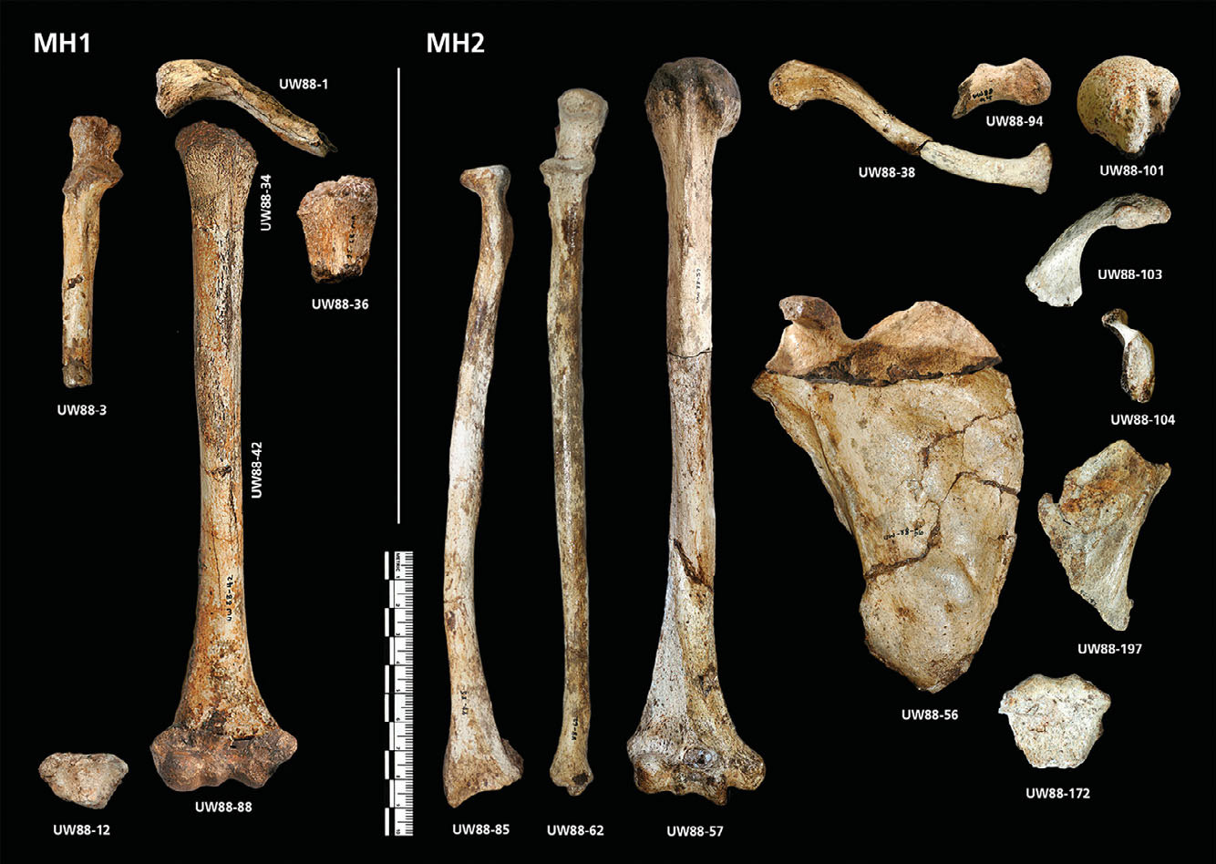
The upper limb elements of Au. sediba specimens (including the designation of the skeleton they came from): MH1 right clavicle; right humerus; UW88-36 left humerus; UW88-3 right ulna; and UW88-12 right radial epiphysis. Also MH2: UW88-172 manubrium, UW88-38 right clavicle, UW88-94 left clavicle, UW88-56 right scapula, UW88-103 left scapular acromion, UW88-104 left scapular glenoid fossa, UW88-197 left scapular body fragment, UW88-57 right humerus, UW88-101 left humerus, UW88-62 right ulna, and UW88-85 right radius. (This image specifically relates to the paper in the journal Science by Churchill et al.)
Comparing clavicles
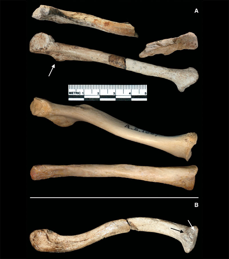
Clavicular morphology in Au. sediba, the chimpanzee (Pan troglodytes), and Homo sapiens from top to bottom (A). The inferior perspective of the clavicle of Au. sediba.
Rib shape
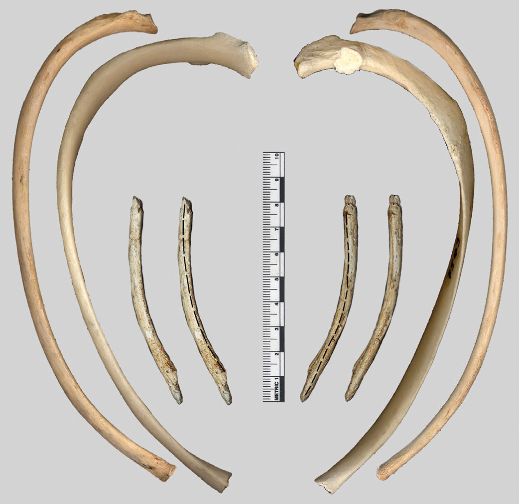
Ninth rib morphology in Au. sediba, the chimpanzee (Pan troglodytes), and Homo sapiens, shown in superior view on the left, inferior view on the right. (This image specifically relates to the Science paper by Schmid et al.)
Skinny chested
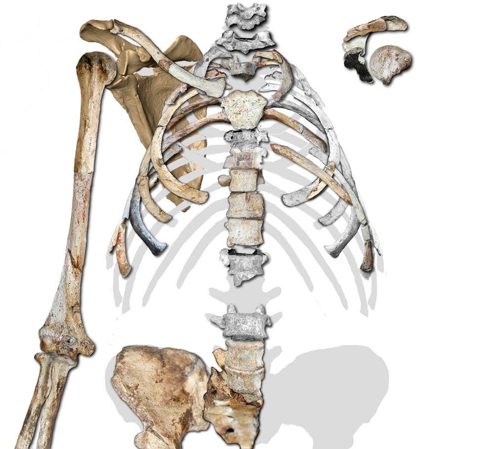
Two-dimensional reconstruction of the upper body of Australopithecus sediba, showing the conical, apelike shape of the upper thorax, different from the broad, cylindrical chest seen in humans. The reconstruction also shows the species' high and lateral position of the shoulder. (This image specifically relates to the Science paper by Schmid et al.)
Sign up for the Live Science daily newsletter now
Get the world’s most fascinating discoveries delivered straight to your inbox.
Lower rib cage
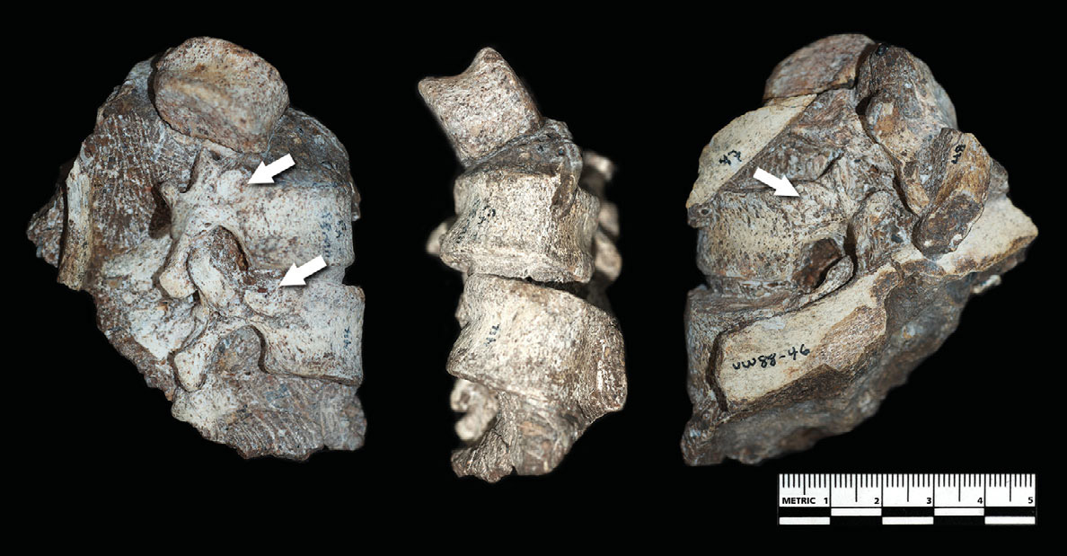
Lower thoracic block of the female Au. sediba (MH2). The less well-preserved lower rib cage fossils were more humanlike, something that might have helped accommodate its strange form of somewhat pigeon-toed walking just as its odd lower back did.
Flexible backs
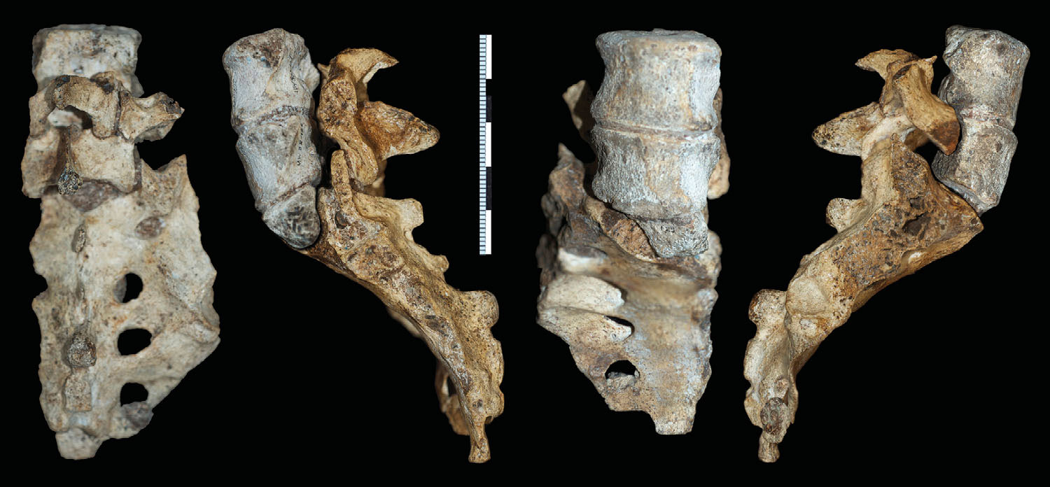
Analysis of its spine revealed Au. sediba had a humanlike curvature of the lower back. However, its lower back was longer and more flexible than modern humans, and more like primitive, extinct members of Homo.
Here, the penultimate (L4) and ultimate (L5) lumbar vertebrae and sacrum of the female skeleton (MH2). (This image specifically relates to the Science paper by Williams et al.)
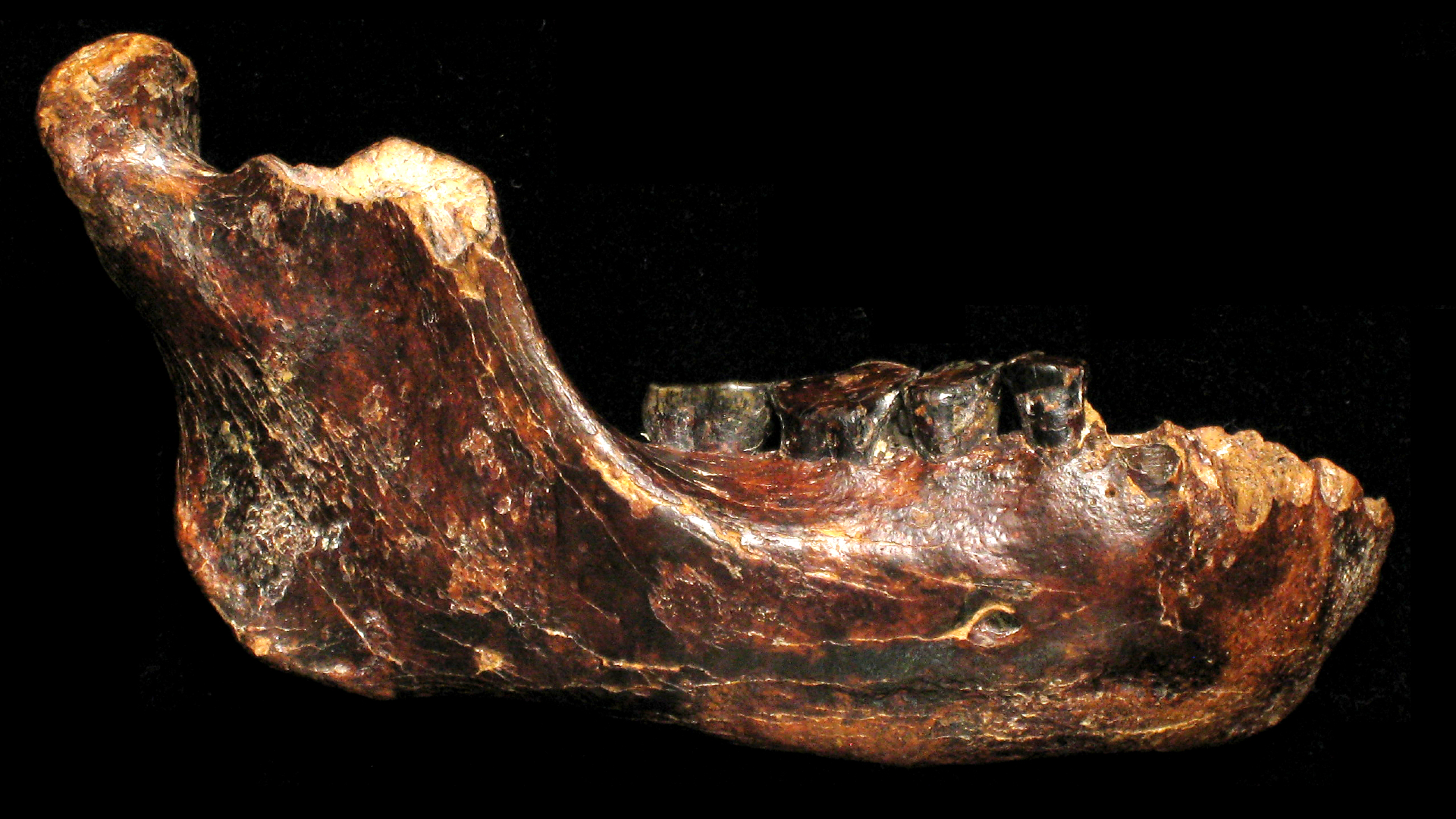
Lower jaw
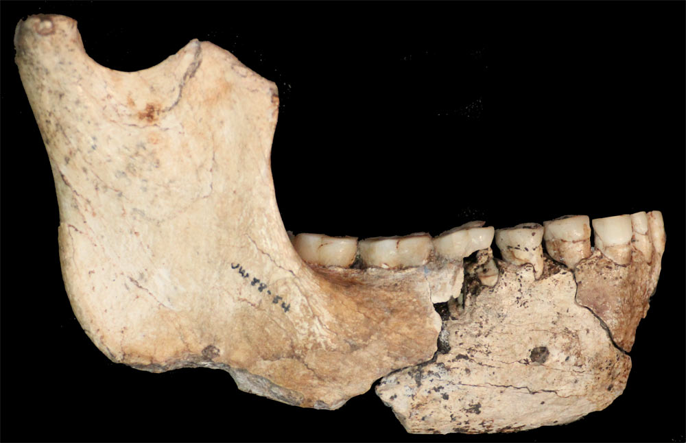
Lower jaw of an adult, female individual of Australopithecus sediba that demonstrates this species differs substantially from other early human ancestors in both size and shape. (This image specifically relates to the Science paper by de Ruiter et al.)
Teeth

The right maxillary (left) and right mandibular dentition of the Au. sediba younger male skeleton. (This image specifically relates to the Science paper by Irish et al.)
