Multiple Myeloma Detection Better with CT Scans

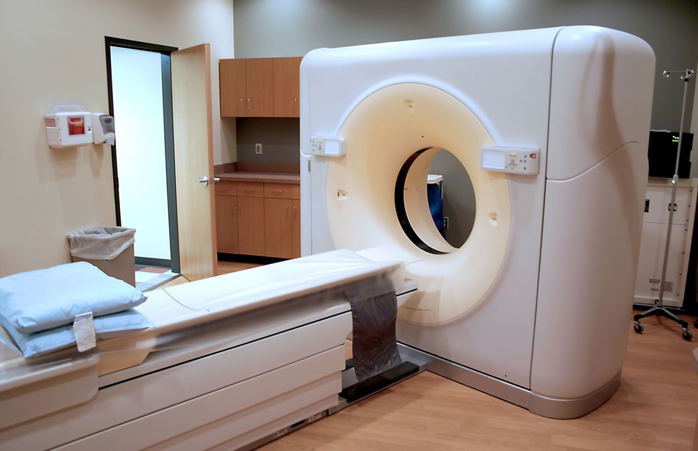
A new study of finds low-dose, whole body CT scans are nearly four times better at detecting multiple myeloma than radiographic skeletal survey, which is currently the standard approach in the United States.
Multiple myeloma is a cancer of plasma cells, a type of white blood cells in bone marrow. The plasma cells normally make antibodies that fight infections. Multiple myeloma symptoms can include fatigue, fractures or damage to bones, kidney failure, and problems with the immune system.
The study, conducted at the University of Maryland in Baltimore, included 51 patients who had both a radiographic skeletal survey as well as a low dose whole body CT examination. The total number of lesions detected in these patients with low dose whole body CT was 968 versus 248 detected by radiographic skeletal survey, said Kelechi Princewill, MD, the lead author of the study.
"The stage of disease determines treatment, and the study found that in 31 patients, the stage of disease would have been different with low dose whole body CT. Thirteen patients would have been upstaged from stage I to stage II; nine patients would have been upstaged from stage I to stage III and nine patients would have been upstaged from stage II to III based on additional lesions detected on the low dose whole body CT," said Dr. Princewill.
Low dose whole body CT was significantly better than radiographic skeletal survey in detecting lesions in the spine, ribs, sternum and flat bones, added Dr. Princewill.
The use of low dose whole body CT is accepted in Europe as an accurate alternative to radiographic skeletal survey for detecting bone lesions in these patients, said Dr. Princewill. A concern about radiation dose may be one of the reasons why it is not widely accepted in the U.S., he said.
"Our study employed a low dose protocol, with an average recorded CT dose of 4.1 mSv. That compares to 1.8 mSv for the radiographic skeletal survey. Using modified protocols and exposure parameters, we were able to significantly reduce the radiation doses to our patients without significantly compromising the image quality required to detect myeloma lesions. The average CT dose used in our study was approximately nine times lower than doses used in the acquisition of standard body CT studies," Dr. Princewill said.
Sign up for the Live Science daily newsletter now
Get the world’s most fascinating discoveries delivered straight to your inbox.
The study is being presented today (May 2) at the American Roentgen Ray Society annual meeting in Vancouver, Canada.


