Magnificent Microphotography: 50 Tiny Wonders
Alien Life or Sparkly Decor?
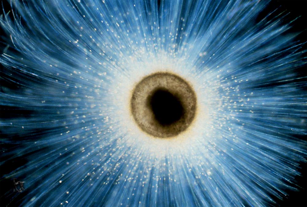
Alien life? An odd extrasolar planet? Maybe an eyeball?
Perhaps just as exotic, this planktonic foraminifera called Orbulina universa evolved about 13 million years ago. The single-celled, shelled organism was captured by scuba divers from surface waters off Santa Catalina Island, Calif.
Living inside the calcite spines of this creature are other simple organisms called dinoflagellates; the dinoflagellates have formed a partnership with the foraminifera, using photosynthesis to produce foods while living in the calcium-rich spines.
At the end of their four-week life cycle, the shells from both protists (foraminifera and dinoflagellate) sink to the seafloor where they become part of the microfossil assemblage in deep-sea sediments. The geochemical compositions of such shells are used to reconstruct past ocean changes. Researchers reported in the Jan. 26 issue of the journal Science Express the importance of such geochemistry; they reported that lithium isotopes in sea sediments reflect several intense episodes of mountain-building and other major geologic events in the last 60 million years.
A Mouse's Hair Trigger
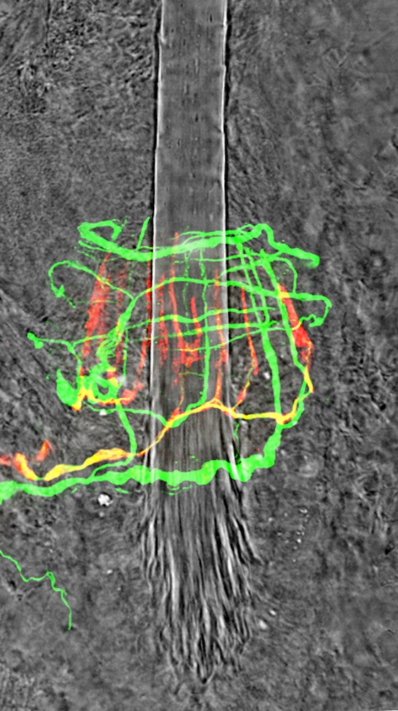
The lightest touch can trigger sensation thanks to these colorful filaments. This image is of nerve fibers wrapped around the hair follicle of a mouse. Run your finger ever-so-lightly across your arm hair: That tickle you feel is the result of nerves like this that detect minute changes in the position of a hair.
New research on sensory signals finds that a protein crucial to eye development is also important for the ability of both humans and mice to sense vibrations. The c-Maf protein is known for its importance in proper eye development; when something goes wrong with c-Maf, cataracts result. It turns out that when c-Maf mutates, Pacinian corpuscles, a kind of touch receptor specialized to detect fast vibrations, also atrophy. Humans have Pacinian corpuscles in our fingertips, meaning that one messed-up protein can damage multiple senses.
Inflamed Beauty
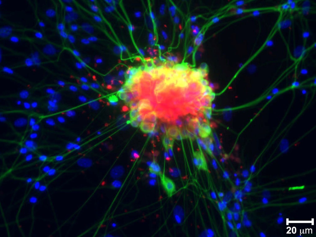
In brilliant blues and greens, this image reveals a tangle of spinal nerve cells, with neurons in green and supportive glial cells all around. Cell nuclei are shown in blue. The red marks the expression of a gene called COX-2, which is stimulated by inflammation.
Vanderbilt University researcher Lawrence Marnett and his colleagues have discovered that COX-2 metabolizes endocannabinoids, which are naturally-occurring painkillers in the body that activate the same brain receptors as marijuana. Intriguingly, certain forms of ibuprofen and other non-steroidal anti-inflammatory drugs (NSAIDs) block this metabolism, the researchers find. That means the pain-killing endocannabinoids stick around longer, partially explaining why popping an Advil can kill a headache.
The Claw
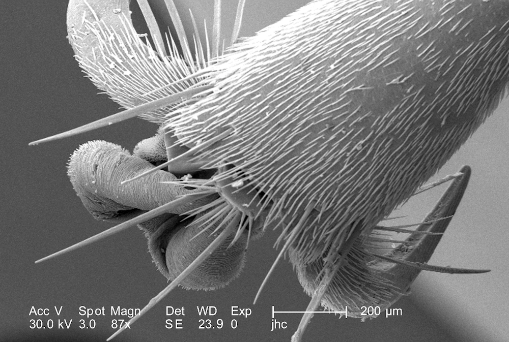
Fancy a handshake with a hornet? This spiky appendage is the foot of an unidentified hornet found in Decatur, Ga. Magnified 87 times, this image is of the insect's "pretarsus," or the tip of one of its six legs. The sucker-like pad in the middle of the hornet's claw is the arolium, and the hair-like projections all over the leg are called setae. This image was taken with a scanning electron microscope in 2007.
Unwelcome Visitor
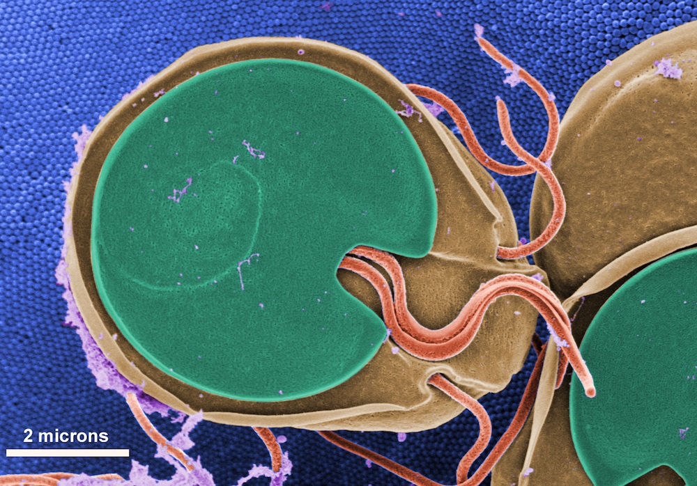
If this fellow is in your well water, don't quench your thirst. This is a scanning electron microscope image of Giardia muris, a protozoan parasite that causes nasty diarrhea when it infects the intestines of its hosts.
Giardia has two phases in its life cycle: the cyst, a dormant phase, and the active trophozoite phase, seen here. People can contract the parasite by drinking water contaminated with cysts; from there, the parasite becomes active, with very unpleasant digestive results. Anti-parasite medication can help fight off these fierce freeloaders, which attach to the intestine lining (seen here in blue). The worm-like flagella seen in this image allow the trophozoites to swim freely in the host's gut.
The Parasite and the Protector
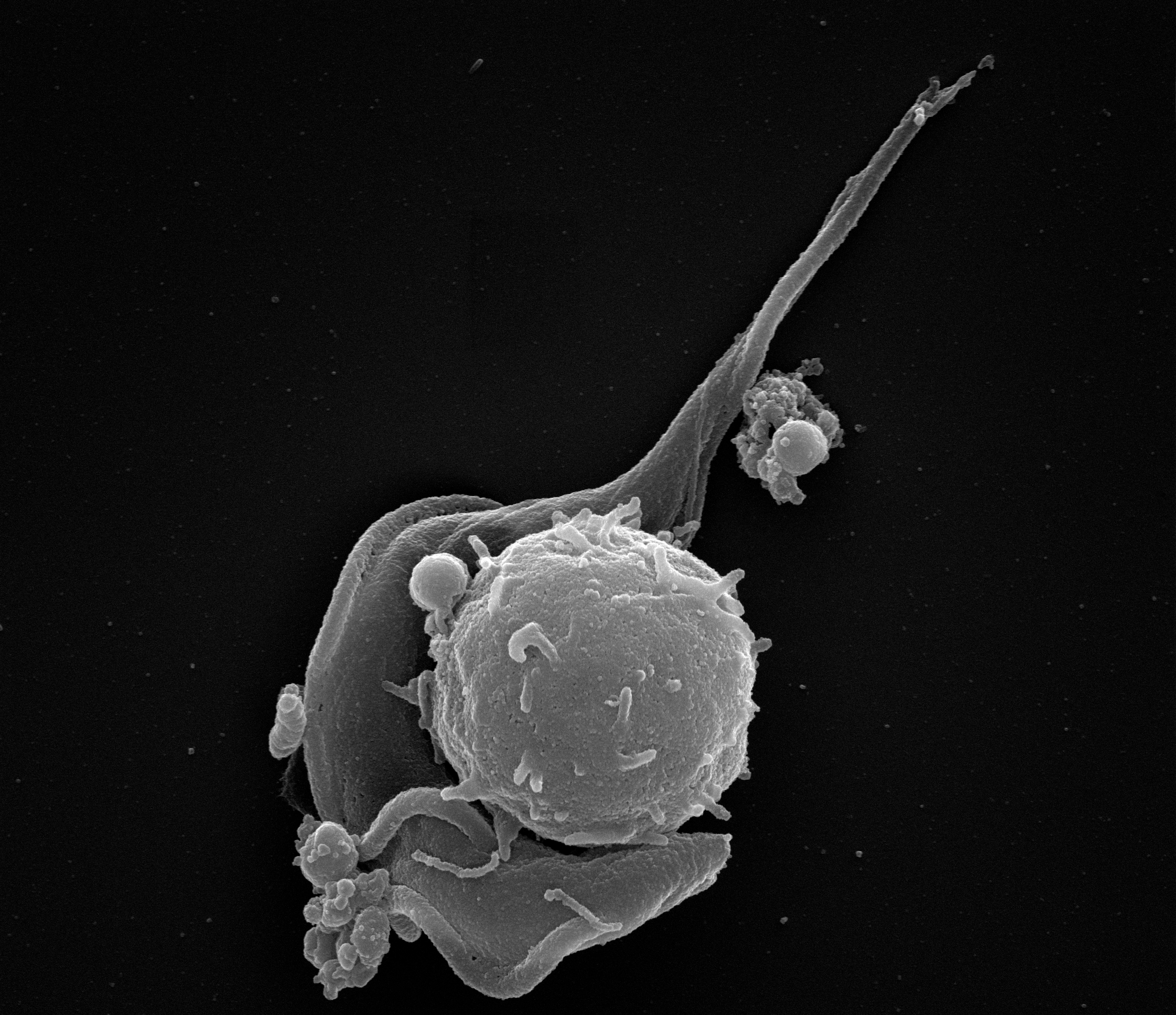
An immune cell tangles with a protozoan parasite in a life-or-death struggle. The ribbon-like parasite is Trypanosoma brucei, a microscopic menace that causes African sleeping sickness. The parasite is transmitted by the bite of the tsetse fly. New research, published June 14, 2012 online by the journal Science, finds that once in the body, this parasite is well-adapted to give the immune system the slip. By releasing certain messenger chemicals, the parasite can shut down the anti-trypanosome proteins in immune cells.
The findings are important, given that at least 7,000 people per year in sub-Saharan Africa contract sleeping sickness, according to the World Health Organization. As the parasite infiltrates the brain, symptoms include disturbed sleep, confusion and poor coordination. If caught early, African sleeping sickness is treatable; left untreated, it is almost always fatal.
What in the World?
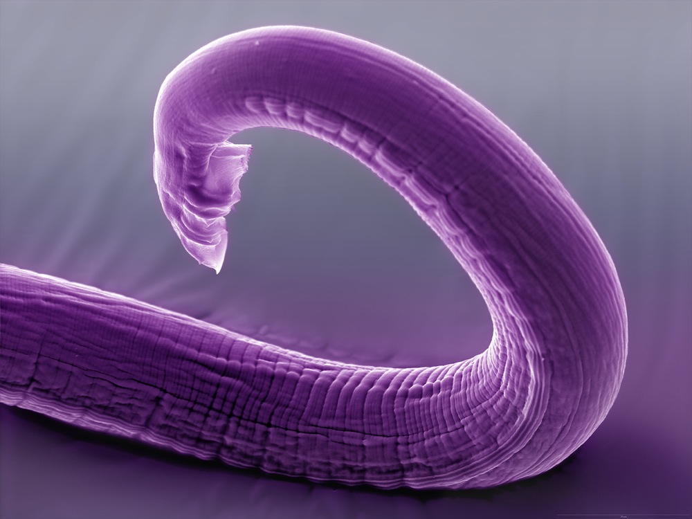
Any guesses as to what this unusual tendril might be? Squid arm? Elephant trunk? Scroll down for the answer …
You're looking at the rear end of a tiny worm called Caenorhabditis elegans, one of the most common lab animals in science. These little soil-living nematodes are only about 0.04 inches (1 millimeter) long. They're handy for scientists because they're easy to analyze genetically and simple to keep alive in the laboratory. C. elegans can even survive being frozen and thawed, making long-term storage easy.
This image comes courtesy a recent study published July 27 in the journal Science. Researchers mapped the neural connections in the nervous system of the C. elegans posterior, revealing the sexual circuits that play an important role in mating. The nerves of a worm's rear end may seem like an odd topic of study, but scientists believe that tracing these simple circuits will help them understand how the more complex neural circuits of humans and other mammals work.
Get the world’s most fascinating discoveries delivered straight to your inbox.
The (Tiny) Face of a Killer
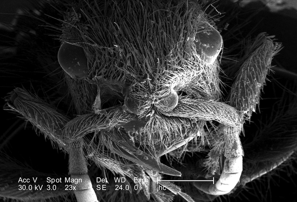
The visage of a tiny velvet ant peers up in this scanning electron microscope image magnified 23 times. This tiny creature, genus Dasymutilla is not actually an ant at all, but a wasp. She (this is a female) boasts a nasty sting, especially if you're another wasp or bee. In order to reproduce, velvet ants lay their eggs inside the larvae of wasps and bees. When the eggs hatch, they feed on the still-living but paralyzed larvae that house them.
There's Hair Where!?
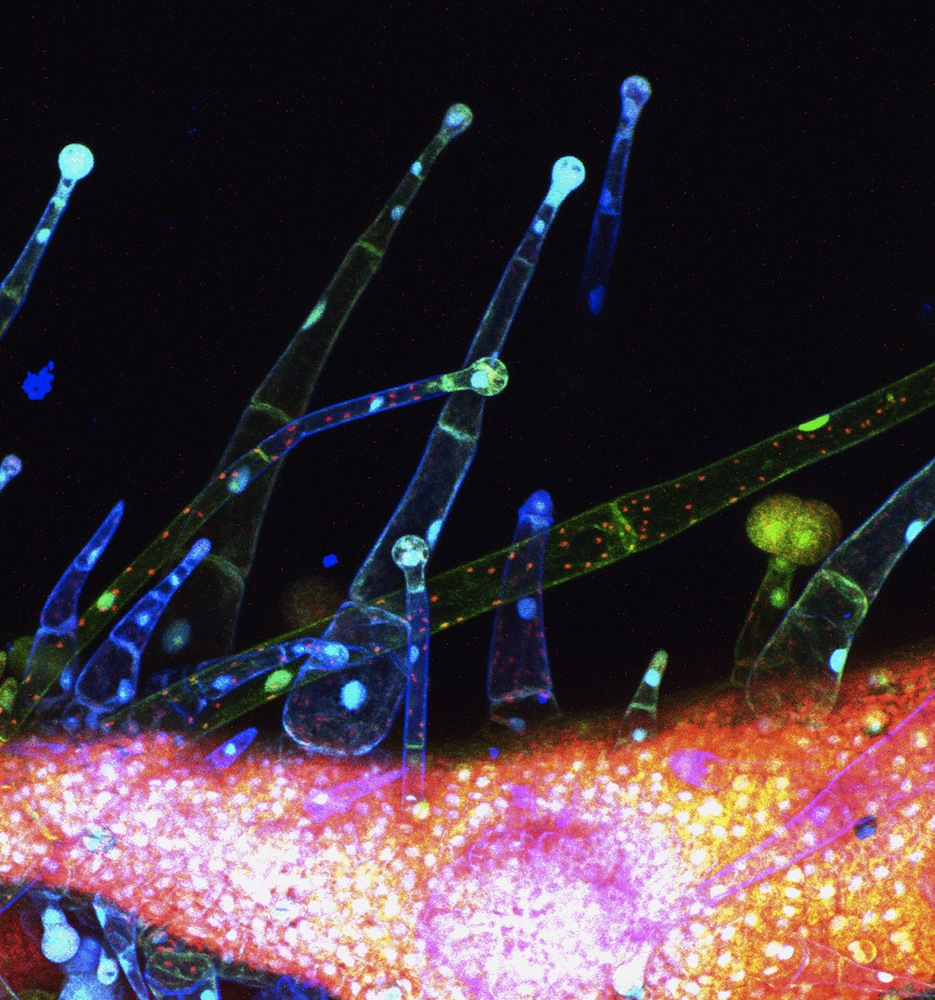
These odd appendages may look alien, but they're definitely terrestrial. In fact, they're practically mundane. These are trichomes, hair-like projections found on plants — in this case, a tomato plant. Researchers at Michigan State University have discovered a gene that allows these trichomes to make acyl sugars, compounds that protect the plants against pests, in cultivated tomatoes. As it turns out, cultivated tomatoes aren't as prolific as wild ones at making acyl sugars, meaning they're more vulnerable to insects and other critters that like to chow down on their leaves. The findings could help researchers engineer heartier tomatoes, the scientists reported in the Proceedings of the National Academy of Sciences.
Winter's Beauty
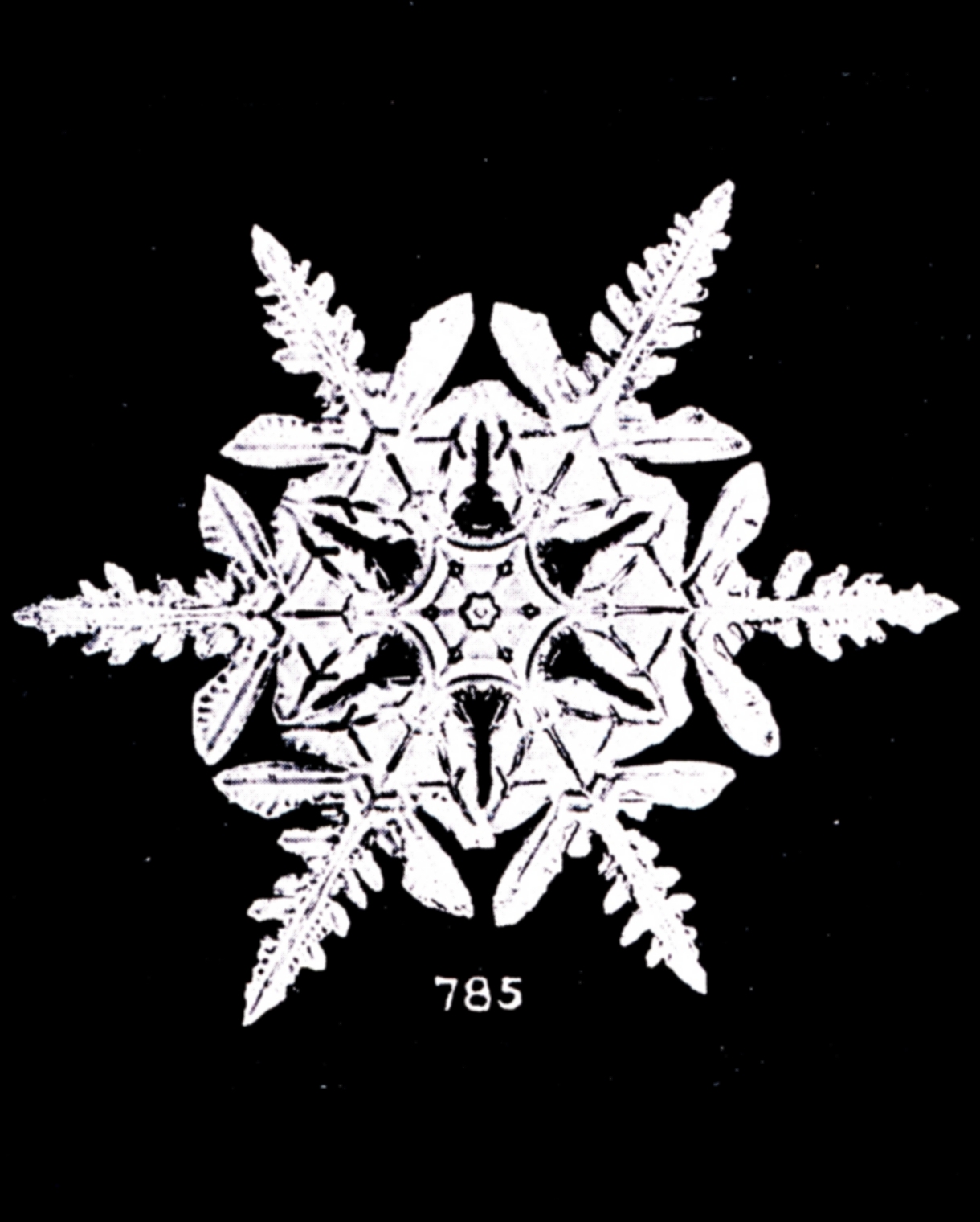
At the winter solstice, the darkest day of the year, it can be tempting to long for warm breezes and green leaves. But winter has its beauty too. This single snowflake was captured on film in 1902 by Wilson Bentley. Known as the "snowflake man," Bentley took over 5,000 stunning close-ups of snowflakes in his lifetime. The Vermont man was one of the first people to ever photograph these tiny ice crystals, and the methods he developed are still used today.

Stephanie Pappas is a contributing writer for Live Science, covering topics ranging from geoscience to archaeology to the human brain and behavior. She was previously a senior writer for Live Science but is now a freelancer based in Denver, Colorado, and regularly contributes to Scientific American and The Monitor, the monthly magazine of the American Psychological Association. Stephanie received a bachelor's degree in psychology from the University of South Carolina and a graduate certificate in science communication from the University of California, Santa Cruz.


