In Photos: Inside a Fruit Fly's Brain
Fly experiment
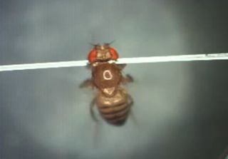
Using lasers, scientists can now surgically blast holes thinner than a human hair in the heads of live fruit flies, allowing researchers to see how the flies' brains work. The researchers also successfully tested this technique on worms, ants and mice.
Prepping for surgery
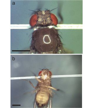
Dorsal and ventral view of fly attached to an optical fiber, which in turn is attached to a silicon fixture. A prominent reflection from the illumination is visible on the thorax as a bright loop. Scale bars are 500 μm.
An overview
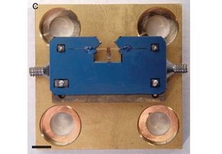
Silicon fixture with fly mounted on brass mount. Scale bars are 5 mm.
Cooling rig
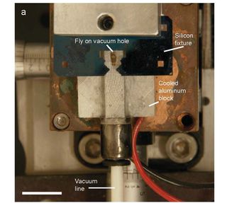
We placed the fly on a cooled aluminum block and attached it to the fiber. The block sits on a thermo-electric cooler, which in turns sits on a copper heat sink. We affixed the latter to a translation stage with six degrees of motion. Scale bars are 10 mm.
Multiple test subjects
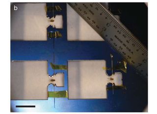
Silicon holder that has four fruit fly mounts precisely etched with 30 mm spacing in both lateral dimensions. Large rectangular holes were etched around the flies to facilitate stimulus delivery. Without these holes, flies could be packed at greater density. Scale bars are 10 mm.
The procedure
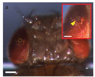
A 35-micron-diameter hole (outlined in yellow) cut in the fly eye using 3,000 laser pulses. The hole extends through the entire fly eye. Scale bars are 100 microns and 50 mcrons in the main panel and inset, respectively.
Another procedure
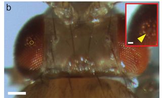
Here, a 20-micron-diameter hole (outlined in yellow) cut in the fruit fly eye using 3,000 laser pulses. The hole is around 250 microns deep and was created without scanning the fly's position. (In comparison, the average human hair is about 100 microns wide.)
Sign up for the Live Science daily newsletter now
Get the world’s most fascinating discoveries delivered straight to your inbox.
Microsurgery
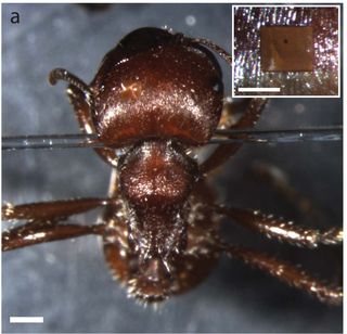
Image shows the red harvester ant (Pogonomyrmex barbatus) after laser microsurgery. The researchers mounted the ant on a 250-micron-diameter fiber (instead of the 125-micron-diameter fibers used for fruit flies). Inset shows the precisely cut edges of the window in the cuticle created using 300 laser pulses. Scale bars are 500 microns for the main panel and 250 microns for the inset.
Clean cut
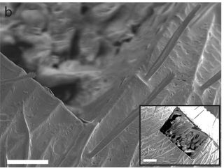
Scanning electron micrograph of an ant after laser microsurgery illustrates the clean edges of the cut. Inset shows the rectangular shape of the hole. Scale bars are 50 μm for the main panel and 100 μm for the inset.
Worms get surgery, too
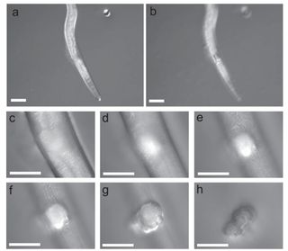
The researchers cut a 12-micron-diameter hole in an anesthetized C. elegans worm using 15 laser pulses. Images show both the plane of incision, (a), and a plane above the incision, (b). Scale bar is 100 microns. (c-h) Images of C. elegans after surgery.
A mouse after microsurgery
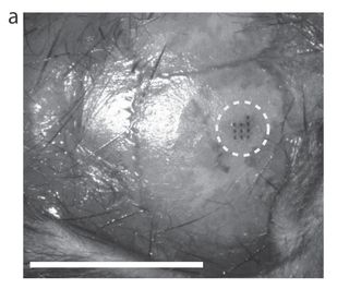
The researchers laser machined a grid of 10 holes ((3 x 3)+1) in the cranium of an anesthetized mouse. Each rectangular hole was 95 microns x 110 microns. Each hole was created in around 3 seconds (600 laser pulses at 200 Hz). Scale bar is 5 mm. Dotted circle highlights the area of the cranium that underwent surgery.

