New Machine Promises Better Images of the Heart
Computed tomography (CT) machines work by sending X-rays around a patient to image cross-sectional slices of the heart. The higher the number of slices, the better the detail.
So a new imager that records 256 slices is expected to be much more effective than the current 64-slice machine.
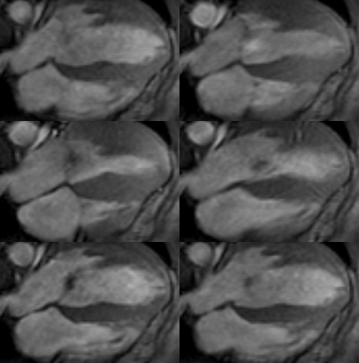
How Heart Attacks Strike The animated MRI image above shows a 4-chamber view of the left ventricle in a patient with aortic insufficiency and left ventricular hypertrophy. Credit: National Heart, Lung and Blood institute
Detailed imaging is important because it can highlight blood vessels and blood flow in the heart and uncover coronary artery disease (CAD). This blockage in the arteries supplying blood to the heart, often causing chest pains and heart attacks, affects about 13 million Americans at any given time and is considered the leading cause of death in this country.
Finding a sick heart
Determining who has heart disease is often a difficult task. Procedures such as exercise and stress tests are often not definitive and patients frequently have to go through invasive procedures that are capable of looking into the artery to find potential blockages.
A new 256-slice scanner, a follow up the 64-slice CT machine, used in conjunction with an injected dye or contrast agent can potentially detect heart defects and coronary artery disease in a single test and in less than half a second while requiring a fraction of the radiation dose that is traditionally needed, according to the developers.
For this reason, the researchers hope this technology can help reduce healthcare costs, by potentially eliminating batteries of unneeded tests in certain cases, says Richard T. Mather a researcher at Toshiba America Medical Systems who will describe the progress of this technology later this month at the annual meeting of the American Association of Physicists in Medicine.
Sign up for the Live Science daily newsletter now
Get the world’s most fascinating discoveries delivered straight to your inbox.
Clear picture
Although the technology is simple, the difficulty of obtaining heart pictures is in its beating. The heart beats so fast and moves so much that pictures turn out blurry. But CT machines gate the X-ray scans to the individual’s heartbeat via cardiograms, a graphical recording of the cardiac cycle produced by an electrocardiograph.
In this way, the machine takes an X-ray image only when the heart muscle is relaxed, eliminating motion. Often times, the heart rate needs to be slowed down in order to obtain the images.
"Heart rate is a big issue with coronary CT imaging," said Jamshid Shirani, head of the Geisinger Medical Center’s cardiology fellowship program, who is not involved with the new technology. "A lot of time and effort is spent to bring the heart rate down to a slow steady rate with medications called beta-blockers and sometimes with other medications such as calcium channel blockers."
Beta-blockers hinder the effects of the hormone and neurotransmitter known as norepinephrine, Blocking norepinephrine slows down the nerve impulses that travel through the heart. The heart then needs les oxygen and blood and starts to pump slower.
Similarly, calcium channel blockers affect the movement of calcium into the heart and blood vessels and reduce the heart's workload and pumping strength.
"There has been [a] rapid evolution of scanners in the last few years and the 256 slicer is the newest kid on the block," Shirani told LiveScience. "The 256 slicer will allow imaging at higher heart rates. There is uncertainty about any reduction in radiation dose and I do not believe at this point that there is any data showing reduction of radiation dose with this new system."
The 256-slice scanner is scheduled for clinical introduction within the next 2 to 3 years.

