3D Images: Exploring the Human Brain
Exploring the human brain
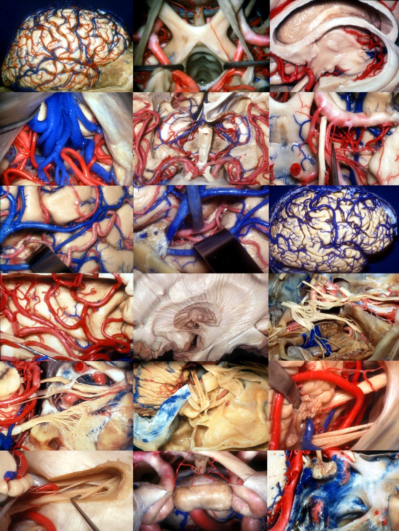
Dr. Albert Rhoton of the University of Florida has collected an unparalleled library of brain-anatomy images. Available in 3D online through iTunes University, the images allow surgeons to view delicate brain structures at precise angles. Bright blue-and-red dyes make blood vessels visible, so surgeons can better plan delicate surgical approaches. Explore the geography of the human brain through this collection of Dr. Rhoton's images.
Right Brain
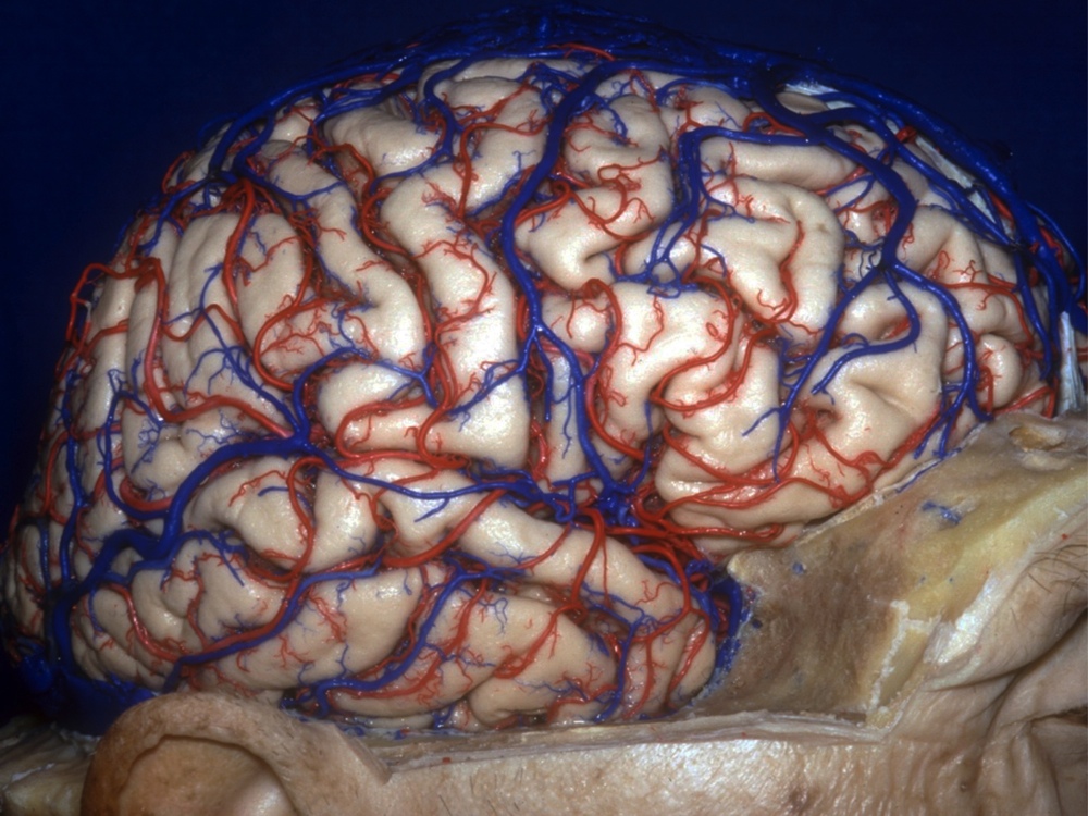
Looking on from the side (laterally), this image shows the right cerebral hemisphere. The cerebrum — site of language, memory and sensory processing — divides down the middle into two sections. Despite the claims of pop psychology, the right hemisphere is not especially creative, nor is the left brain inherently more logical. Sensory information from each side of the body, however, does travel to the opposite side of the brain.
Brain Cushion
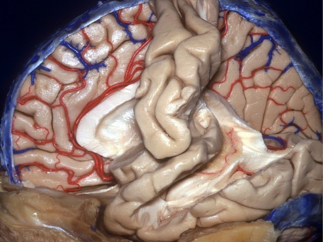
Here, the left hemisphere has been almost entirely removed, revealing the surface of the right brain where it meets the cerebral divide (a "medial view"). The arteries and veins can be seen snaking through the brain tissue. The large, white, horn-shaped structure in the middle is a lateral ventricle, a cavity filled with cerebrospinal fluid that cushions the brain.
Eye Line
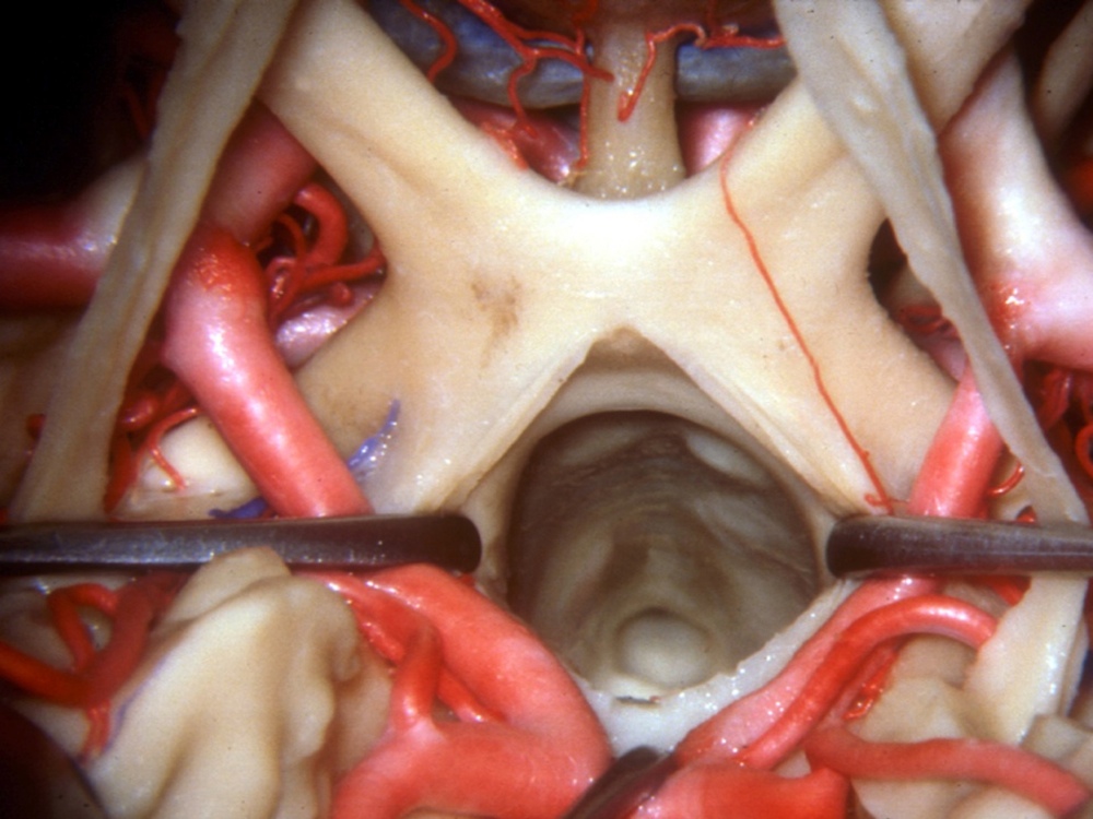
Peer down into the optic chiasm — a site that plays a major role in a person's ability to peer in the first place. The chiasm marks the point where some of the optic nerves crisscross on their way from the eyes into the brain. Images that strike the nasal side of each retina cross over to the opposite side of the brain.
Below the Cerebellum
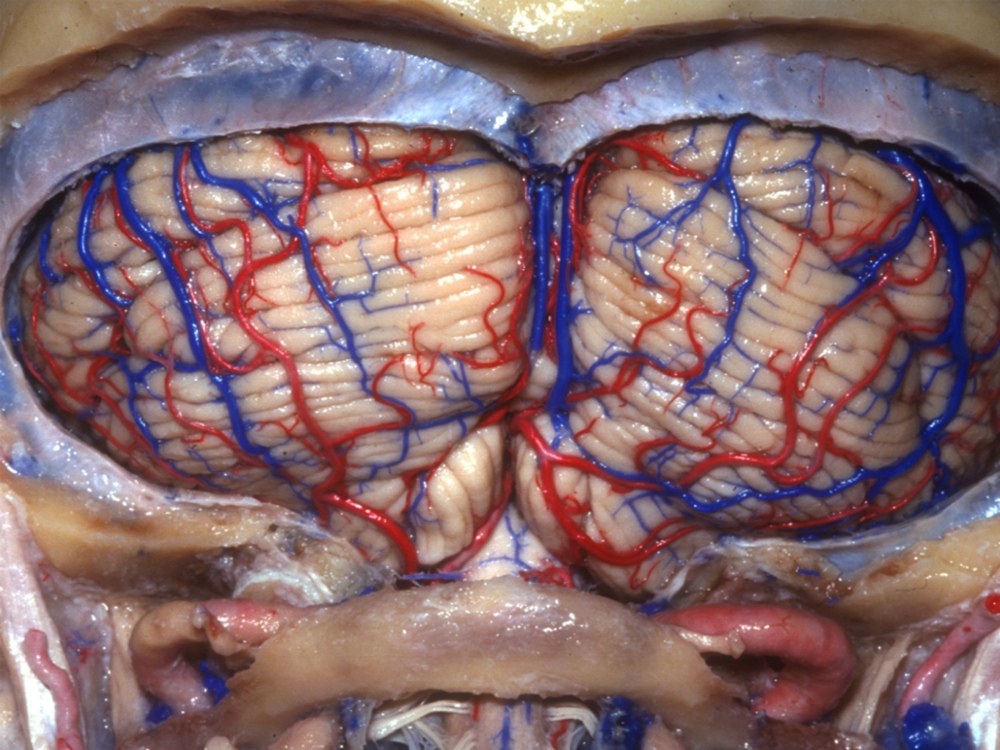
The cerebellum, a brain region important for motor control, looks like a separate organ, stuck onto the brain below the two hemispheres. This image shows the "suboccipital surface" of the cerebellum — that is, approached from below. The cerebellum doesn't initiate movement, but this brain region governs proper coordination and timing.
Cerebellum Base
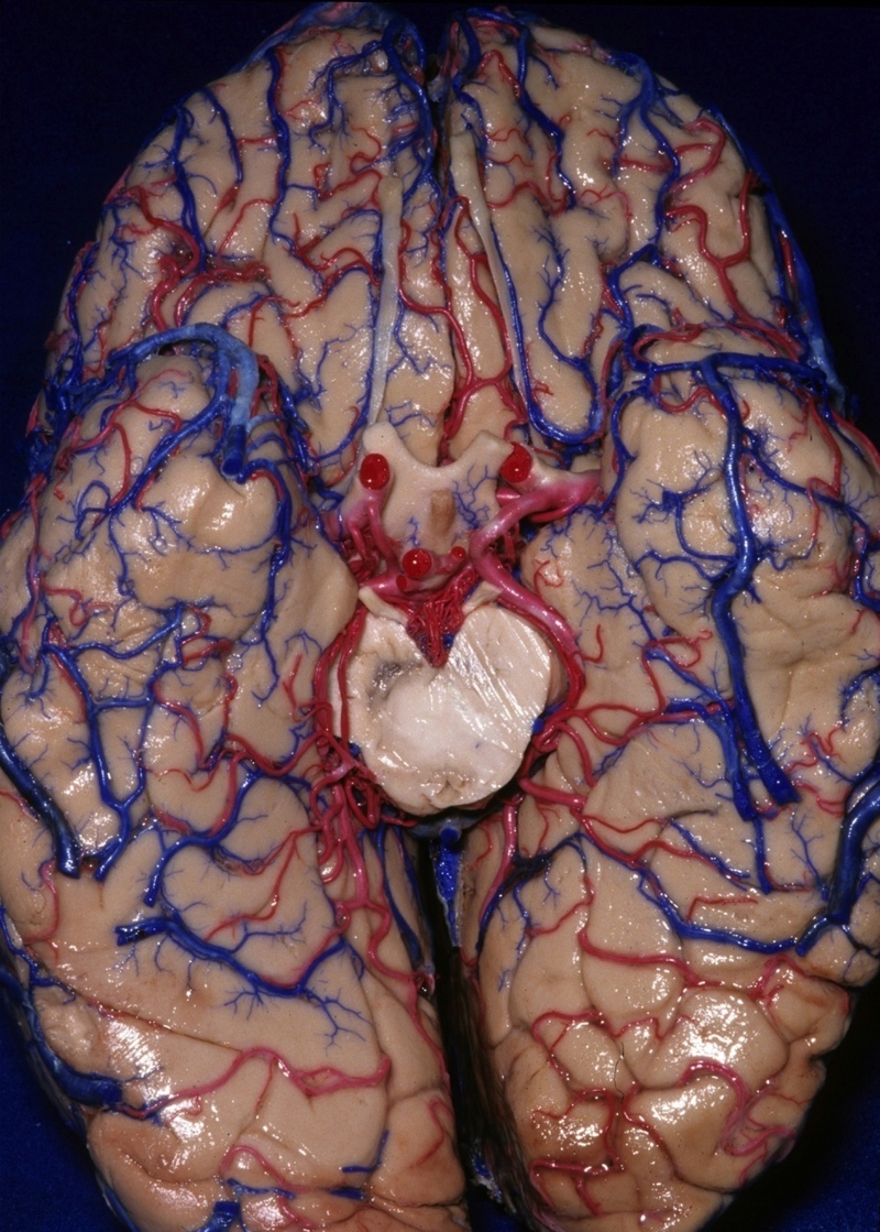
Here, the cerebellum returns, but viewed from its "base," or where it attaches to the rest of the brain (a "basal" view). A tough layer of tissue called dura mater separates the cerebellum from the cerebrum. However, the cerebellum still gets information from other parts of the brain, via connections to part of the brainstem called the pons.
Spinal Cord
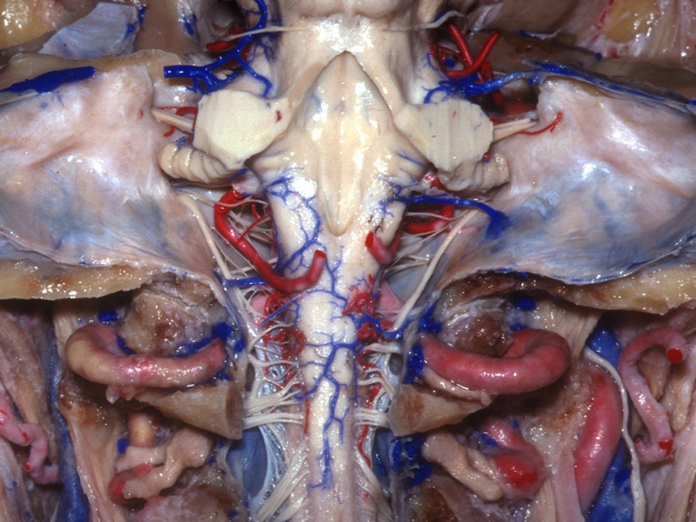
With the cerebellum removed, the top of the spinal cord appears. This is a "posterior" view, meaning it looks toward the spinal cord from behind the body. The spinal cord attaches to a region of the brain called the medulla oblongata, a portion of the brain stem responsible for involuntary functions like breathing.
Sign up for the Live Science daily newsletter now
Get the world’s most fascinating discoveries delivered straight to your inbox.
Big Brain Vein
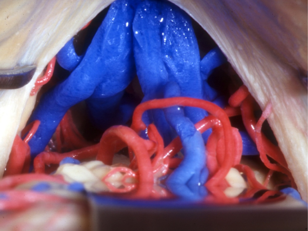
The big blue structure here (dyed by Rhoton for easy viewing) shows where the great cerebral vein drains blood from the cerebrum. This vein also goes by "the Vein of Galen," named for the ancient Greek doctor, Galen, who discovered it. The pineal gland, which produces a hormone affecting sleep patterns, is also visible here.
Half a Brain
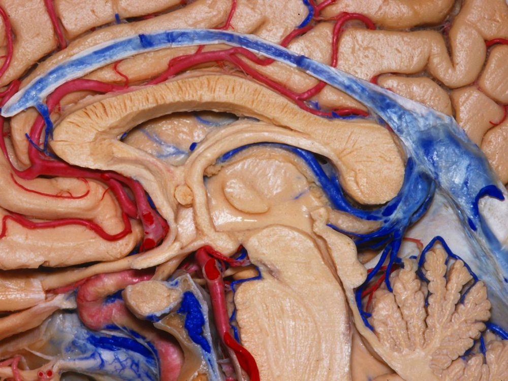
Here, the brain has been neatly sliced in half — a "midsaggittal section." This piece of the section highlights the pituitary gland, the little round piece surrounded by blood vessels, located just behind the nose and below a region of the brain called the hypothalamus (lower left). Called the "master gland," the pituitary releases hormones that influence other glands.
Brain Stem
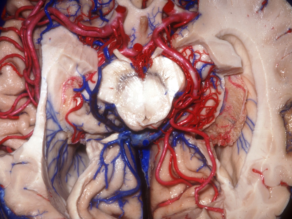
The lateral ventricles (cavities providing cushioning) and other structures surround the brain stem in this image. The brain stem controls basic body functions, such as breathing and blood pressure. It also serves as an important hub: The neurons responsible for transmitting sensory and motor (muscle-movement) information between the brain and the body travel through the brain stem.
Nerve Cluster
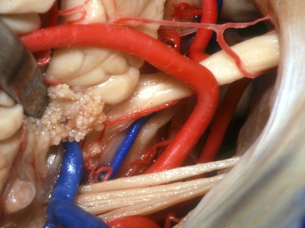
This cluster of nerves and arteries meets at the "cerebellopontine angle," a juncture between the cerebellum and the pons. Part of the brain stem, the pons mediates all message transfers between the cerebellum and the rest of the brain. Brain surgeries, like those performed by Rhoton, must take great care to avoid damage to such nerves and blood vessels.

Michael Dhar is a science editor and writer based in Chicago. He has an MS in bioinformatics from NYU Tandon School of Engineering, an MA in English literature from Columbia University and a BA in English from the University of Iowa. He has written about health and science for Live Science, Scientific American, Space.com, The Fix, Earth.com and others and has edited for the American Medical Association and other organizations.
