A new printing method inspired by kids' stamps could be used to create live cells of almost any shape or configuration.
The technique, called BlocC printing, could be used to recreate networks of brain cells in a petri dish or complicated immune-system interactions, according to the study detailing the method, which was published today (Feb. 10) in the journal Proceedings of the National Academy of Sciences.
And unlike past cell-printing methods, "the major improvement is that cells printed by BlocC printing are alive — close to 100 percent viability," said study co-author Lidong Qin, a nanomedicine researcher at Houston Methodist Research Institute.
Printing cells
Scientists have used printing methods to build bone and eye cells, and even print embryonic stem cells. [See Images of the Printed Cells]
Some scientists even hope 3D methods could eventually be used to print whole organs on demand or make such realistic cell-cultures that they render animal testing obsolete.
But most of these methods relied on a variant of ink-jet printing, which can create high shear forces as nozzles spit out cells, meaning only some of the printed cells survive.
Sign up for the Live Science daily newsletter now
Get the world’s most fascinating discoveries delivered straight to your inbox.
"We were sick of using ink-jet printing and started to think of other approaches to prepare a cell pattern," Qin told Live Science in an email.
Stamping process
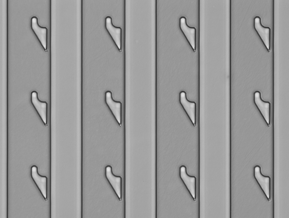
So the team drew inspiration from watching little kids play with rubber stamps. The researchers created silicone molds and guided cells into the mold using tiny, hooklike traps. The cells filter down a column and move past cells that are trapped to fill the next space in the mold. When the mold is removed, the cells are left behind in the exact configuration of the mold. In basic principle, the system isn't much different from ancient Chinese woodblock printing or the big blocks used to print newspapers.
Unlike with the ink-jet printing method, almost all of the cells survived when the researchers used the new technique. But because it only forms 2D, not 3D shapes, the the new technique couldn't be used to print organs, Qin said.
"This is fantastic work," said Ke Xu, a chemist at the University of California, Berkeley who was not involved in the study.
The current new method can deal with many cells of different types — something past techniques could not do, Xu said. That means the system could recreate a realistic, complicated system of multiple cells, better capturing immune-cell interactions, for instance, Xu told Live Science.
One of the most exciting potential applications would be for recreating mini-brain cell networks in the lab. Previous methods often jammed a bunch of cells together, but neurons have branchlike projections, called dendrites, protruding from them, so they need to be spaced more precisely to accurately capture how signals are transmitted between them, Xu said.
Follow Tia Ghose on Twitter and Google+. Follow Live Science @livescience, Facebook & Google+. Original article on Live Science.

Tia is the managing editor and was previously a senior writer for Live Science. Her work has appeared in Scientific American, Wired.com and other outlets. She holds a master's degree in bioengineering from the University of Washington, a graduate certificate in science writing from UC Santa Cruz and a bachelor's degree in mechanical engineering from the University of Texas at Austin. Tia was part of a team at the Milwaukee Journal Sentinel that published the Empty Cradles series on preterm births, which won multiple awards, including the 2012 Casey Medal for Meritorious Journalism.


