Windows to Disease: Eye Vessels Shed Light on Health

Updated on Tuesday, Feb. 18 at 9:10 a.m. ET.
For poets and lovers, the eyes are the windows of the soul. For scientists and doctors, blood vessels at the back of the eye are windows into many diseases.
Blood vessel abnormalities can indicate a variety of serious conditions such as atherosclerosis (hardening of the arteries), heart attacks and strokes. But most vessels are buried beneath skin and other tissues, making them difficult to examine without surgery.
There's one exception — in the eye. Unlike anywhere else in the body, larger vessels on the retina at the back of the eye are directly visible through the pupil, requiring essentially only light and magnifying lenses to view.
These vessels are used to diagnose glaucoma and diabetic eye disease. Because they display characteristic changes in people with high blood pressure , some researchers hope retinal vessels might one day help predict an impending stroke, congestive heart failure or other diseases stemming from dangerously high blood pressure.
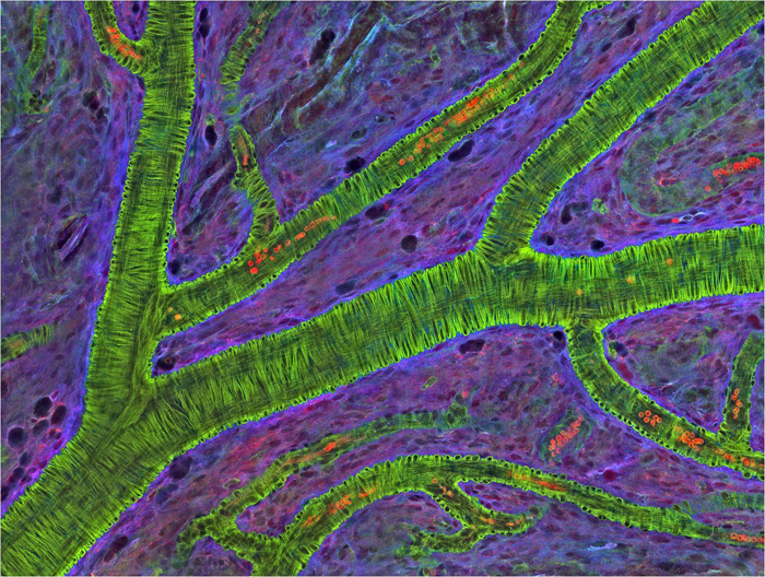
The medical importance of retinal vessels piqued the interest of scientists funded by the National Institutes of Health at the National Center for Microscopy and Imaging Research (NCMIR) at the University of California, San Diego, who captured this micrograph image of mouse retinal vessels.
In the image, the vessels appear green. It's not actually the vessels that are stained green, but rather filaments of a protein called actin that wraps around the vessels. Most of the red blood cells were replaced by fluid as the tissue was prepared for the microscope. The tiny red dots are red blood cells that remain in the vessels.
Get the world’s most fascinating discoveries delivered straight to your inbox.
This image is just one of thousands taken by NCMIR scientists, who develop, test and share high-tech visualization tools and methods to study tissues, cells and molecules. These and other technologies allow researchers to examine life processes in new ways--and also produce stunning scientific images that reveal the beauty and complexity of biology.
The research reported in this article was funded in part under NIH grant GM103412.
Editor's Note: This article has been updated to take out a mention of a forthcoming exhibition of the images, because that exhibition has not been made official to date.
This Inside Life Science article was provided to LiveScience in cooperation with the National Institute of General Medical Sciences, part of the National Institutes of Health.
Learn more:
National Center for Microscopy and Imaging Research
NCMIR Images in the NIGMS Image and Video Gallery
Emerging Technologies Look Deeper into the Eyes Articlefrom NIH's National Eye Institute
Also in this series:
The Amazing World Inside a Human Cell

