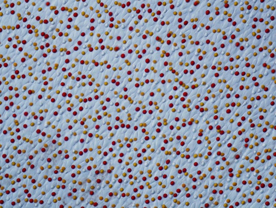
Scattered dots of red and yellow decorate what appears to be an icy, blue terrain in this award-winning microscope photo, which is actually an image of a painted turtle's retina magnified 400 times.
Dr. Joseph Corbo, a researcher at Washington University School of Medicine who studies how retinal photoreceptors interact with the nervous system, captured this remarkable image using the optical microscopy illumination technique known as differential interference contrast microscopy. Cordo used this complex investigative technique—commonly used in science on transparent specimens—to reveal “invisible” features of the painted turtle’s light-sensitive membrane inside the eye.
Differential interference contrastmicroscopy produces a monochromatic shadow-cast image that effectively displays the gradient of optical paths for both high and low spatial frequencies present in the specimen, according to Nikon. Corbo’s image took 2nd prize at the 2013 Nikon's Small World competition. [See the Winning Microscopic Images]
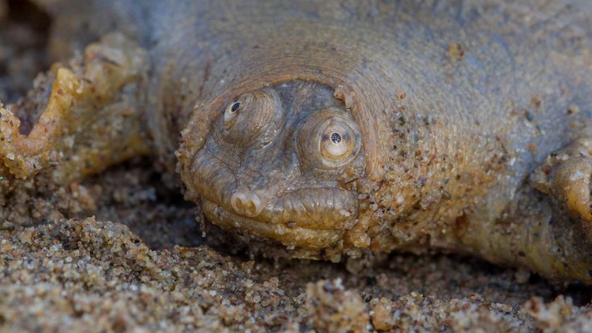
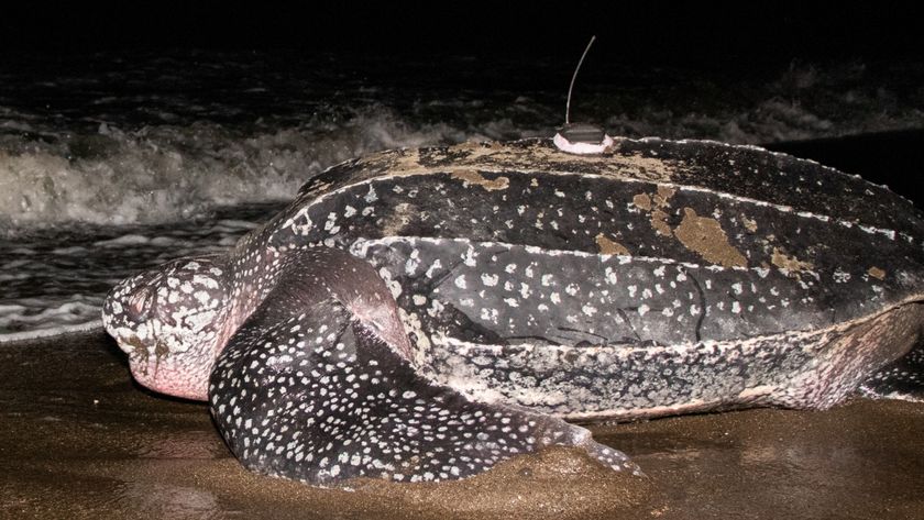
The painted turtle, chrysemys picta, is a fresh-water dwelling reptile found throughout North America.
Follow Live Science @livescience, Facebook & Google+.
Sign up for the Live Science daily newsletter now
Get the world’s most fascinating discoveries delivered straight to your inbox.

