The Diversity of Diatoms

Little, but a big deal
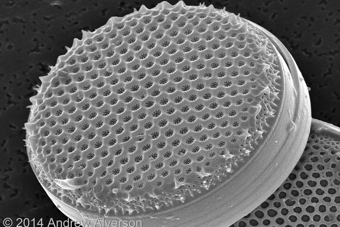
Andrew Alverson is a biologist and expert on diatoms at the University of Arkansas in Fayetteville. He contributed this article to Live Science's Expert Voices: Op-Ed & Insights.
About 20 percent of the oxygen we breathe comes from photosynthesis by marine diatoms — small single-celled algae that play an immense role in keeping the planet's ecosystem working.
Diatoms help mediate carbon and oxygen cycles, are a foundational component of marine food webs, and they process an incredible amount of silica, which comprises about one-quarter of the earth's crust.[The Air You're Breathing? A Diatom Made That]
Andrew Alverson, a biologist and diatom expert at the University of Arkansas in Fayetteville, is studying diatom genomes to answer such questions as how diatoms evolved, how much of their genome they share with bacteria, and how these microorganisms became so well integrated into the global ecosystem.
What follows are ten of the most engaging images of diatoms from Alverson's research.
Amphora
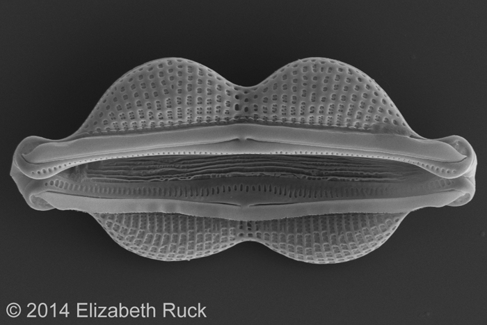
Scanning electron micrograph of the marine diatom, Amphora.
Corethron
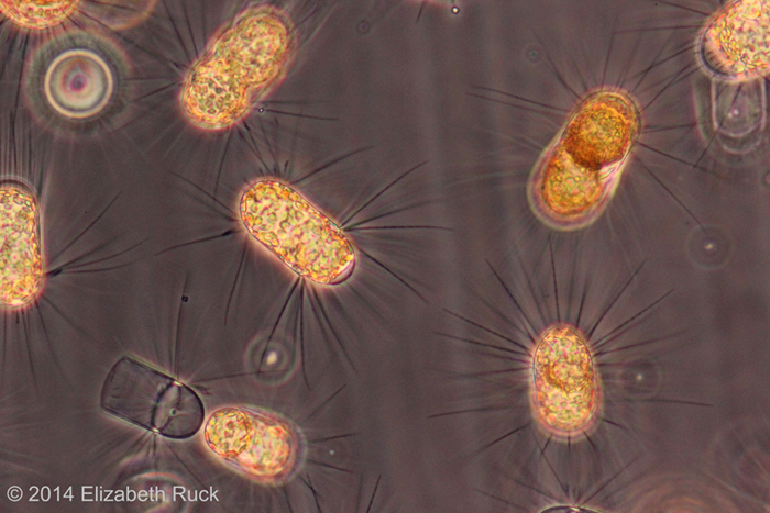
Light micrograph showing the golden-brown plastids and siliceous spines of the marine diatom, Corethron.
Cyclotella quillensis
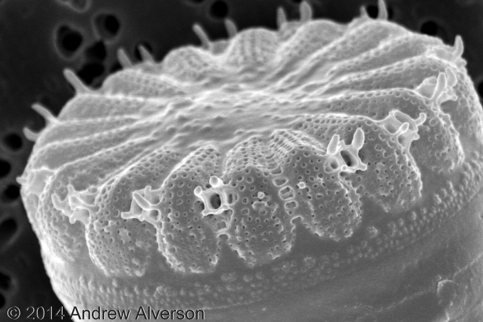
Scanning electron micrograph of the freshwater diatom, Cyclotella quillensis.
Ditylum
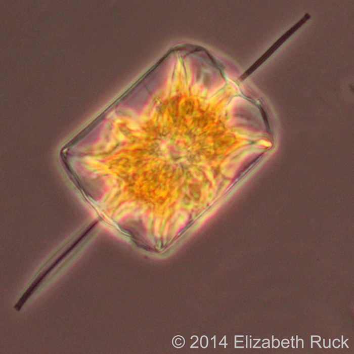
Light micrograph showing the golden-brown plastid of the marine diatom, Ditylum. This is a model diatom for understanding population structure in widely distributed marine phytoplankton species.
Guinardia striata
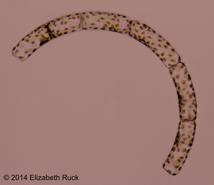
Light micrograph showing a semicircular chain of sibling cells of the marine diatom, Guinardia striata.
Psammodictyon
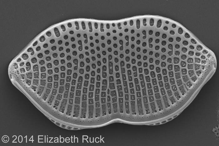
Scanning electron micrograph of the brackish water diatom, Psammodictyon.
Get the world’s most fascinating discoveries delivered straight to your inbox.
Skeletonema
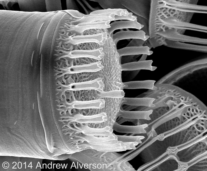
Scanning electron micrograph of the marine diatom, Skeletonema. DNA sequence data have shown that this genus contains many more species than were recognized based solely on their cell wall features.
Thalassiosira nodulolineata

Scanning electron micrograph of the marine diatom, Thalassiosira nodulolineata.
Stephanodiscus
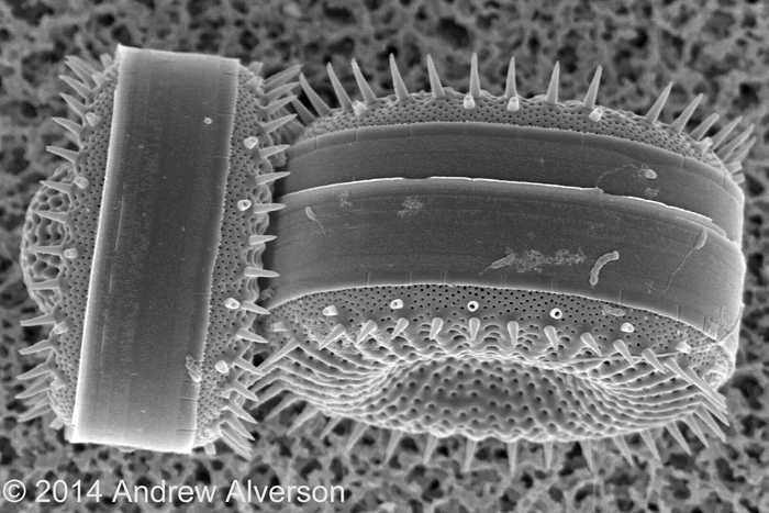
Scanning electron micrograph of the freshwater diatom, Stephanodiscus.
Triceratium
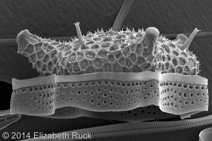
Scanning electron micrograph of the marine diatom, Triceratium.
Follow all of the Expert Voices issues and debates — and become part of the discussion — on Facebook, Twitter and Google +. The views expressed are those of the author and do not necessarily reflect the views of the publisher. This version of the article was originally published on Live Science.


