Artificial Rat Brain Gets Pounded in Name of Science
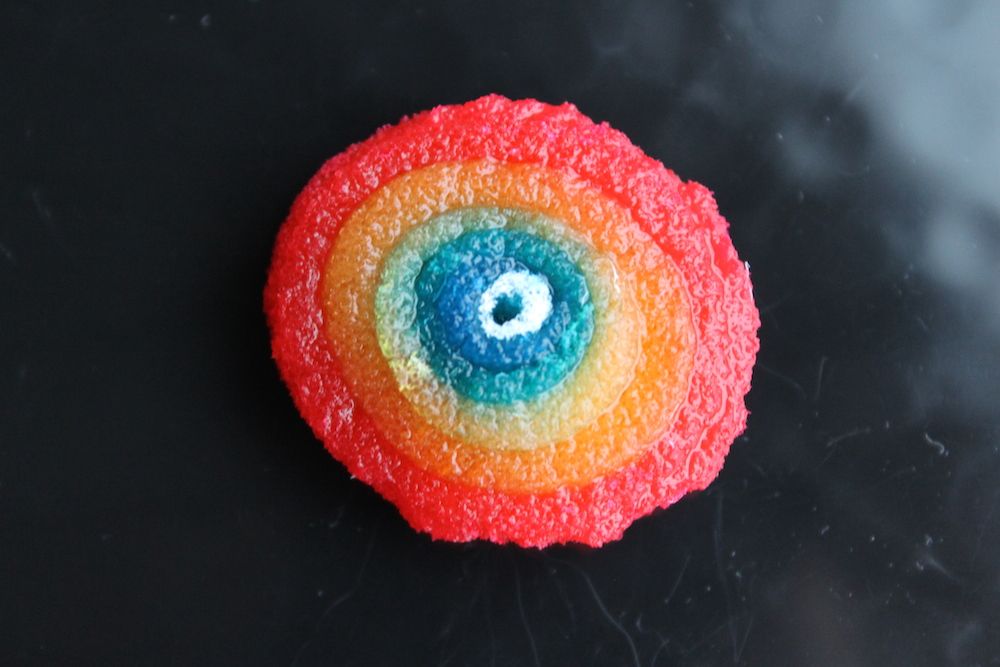
After a new 3D artificial "rat brain" was created in the lab, it got some bizarre treatment: Researchers dropped weights onto the tissue from different heights to see how it might react to a traumatic injury.
The new brainlike tissue is one step toward creating a functioning brain in a petri dish — something that is still a ways off, scientists say. Even so, the simulated brain tissue could be used to study normal brain function, or injured and diseased brains, in order to develop new treatments, researchers say.
The brainlike tissue, which structurally resembled a rat brain, stayed alive for more than two months, and after it was pounded with weights, its neurons showed electrical and chemical activity similar to what is seen in animal studies of traumatic brain injury. [3D Images: Exploring the Human Brain]
"There are few good options for studying the physiology of the living brain, yet this is perhaps one of the biggest areas of unmet clinical need when you consider the need for new options to understand and treat a wide range of neurological disorders associated with the brain," David Kaplan, a biomedical engineer at Tufts University in Boston and lead author of the study published today (Aug. 11) in the journal Proceedings of the National Academy of Sciences, said in a statement.
How to make a brain
The current methods of creating brain tissue in a lab include growing neurons in a 2D mat on a petri dish or in a 3D gel environment.
The 2D petri-dish environment can't replicate the sophisticated 3D structure of gray matter and white matter in a living brain. Gray matter consists of neuron cell bodies, and white matter consists of long-range connections, or axons. Brain injuries and diseases affect these two tissue types differently, the researchers said.
Sign up for the Live Science daily newsletter now
Get the world’s most fascinating discoveries delivered straight to your inbox.
Meanwhile, the 3D gel structures don't survive for very long and don't function the way real brain tissue does because they often lack the complex soup of chemical signals that normally guide the growth and development of brain cells.
In the new study, researchers succeeded in developing a new kind of tissue for modeling the brain, which incorporates both gray matter and white matter. They created concentric doughnut-shaped scaffolds made out of a stiff silk material which they seeded with neurons, and filled them with a softer, collagen-containing gel that encouraged the growth of axons that connect the cells.
Within three days, the axons had grown into the collagen gel; by the second week, they had reached a length of about 0.04 inches (0.09 centimeters). However, "it is unclear what the final length of the axons were and whether axons could grow longer in a larger compartment," the researchers wrote in the study.
Studying brain disease
The researchers measured the health and function of the faux brain tissue over the course of a few months, comparing it with neurons grown in gel alone or in a 2D petri dish. The new tissue survived for at least nine weeks in the lab — much longer than the other kinds of 3D brain tissue, the team reported. The artificial neural tissue also resembles that of a rat's brain, because it had similar mechanical properties, they said.
The development of simulated brains could mean that fewer animal brains would need to be used in studies of brain injury, the researchers said. In addition to addressing such ethical issues, this method would save time, as researchers wouldn't need to dissect and prepare animal tissue for study. Also, because the brainlike tissue survives for months in the lab, researchers could use it to track diseases they wouldn't ordinarily be able to study. They could also use it to study healthy brains, the researchers said.
Follow Tanya Lewis on Twitter and Google+. Follow us @livescience, Facebook & Google+. Original article on Live Science.

