Will Mobile Labs Finally Halt Killer Frog Fungus? (Op-Ed)
Tracie Seimon is a molecular scientist for the Zoological Health Program at the Wildlife Conservation Society (WCS). She is based at the Bronx Zoo in New York City. This article is the first in a series celebrating the contributions of women to the practice of conservation. Seimon contributed this article to Live Science's Expert Voices: Op-Ed & Insights.
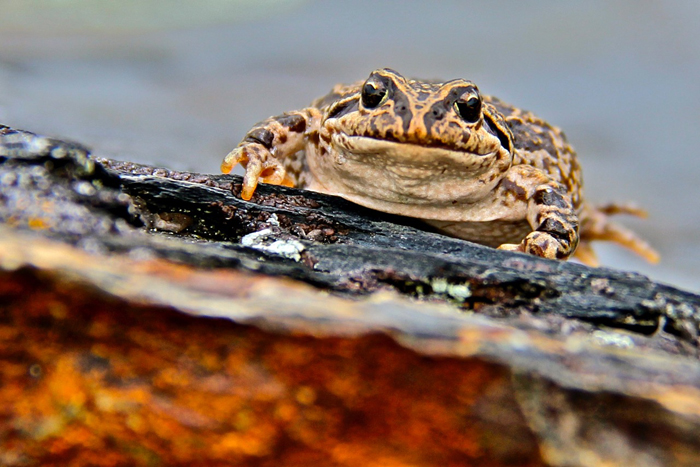
The chytrid fungus is a modern-day scourge of toads, salamanders and frogs around the globe, one of the greatest conservation threats amphibians face. As a waterborne pathogen, the fungus's highly infectious life stage called a zoospore infects amphibian skin, then multiplies. As the disease infects more and more skin cells, the infected animals lose the ability to remain well hydrated and to regulate temperature. Eventually, they lose the ability to breathe.
Some species are resistant to infection; in others, the death toll in a local population can reach 100 percent. While the total number of affected species is unknown, global studies show the fungus is a major factor in the global decline and extinction of amphibian species, with roughly a third of these species threatened throughout the world. [Freaky Frog Photos: A Kaleidoscope of Colors (Gallery)]
As a molecular scientist, my role is to develop or adopt tests that can discover or detect diseases of conservation concern, like the fungus chytridiomycosis. Based at the Bronx Zoo, I usually diagnose diseases in zoo animals, but I also travel to some of the world's most remote protected areas, with some of the world's greatest biodiversity, to sample and test animals for disease-causing microorganisms.
Frog swabbing
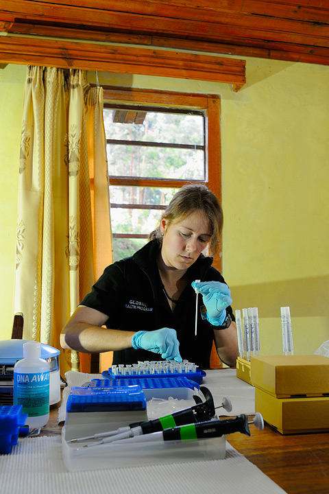
In my travels abroad with my team — to Rwanda, Peru, Myanmar and Uganda, to name a few — one of the first things I pack is my portable freezer. It may be an unusual item to include in one's luggage, but the thermal extremes in the environments where we work range from hot and buggy lowlands to high mountain ice fields to frigid forests. It is critically important that we protect the samples we collect from those challenging environments so we can ensure reliable test results. So the freezer is a must.
We also pack three or four 30-gallon (113 liters), hard plastic cases crammed full of plastic tubes, pipettes, hundreds of pairs of rubber gloves, a polymerase chain reaction (PCR) machine that replicates and measures DNA, a centrifuge that spins samples at 12,000 revolutions per minute, and — my personal favorite — a rubber frog. I use this frog to train people in proper techniques for swabbing and safely collecting samples from amphibian skin for chytrid testing.
Sign up for the Live Science daily newsletter now
Get the world’s most fascinating discoveries delivered straight to your inbox.
It's important to keep a full inventory of all of our equipment, about 300 individual items. If one component is missing, the whole trip is in jeopardy. There are no chemical supply stores I can just pop into while in a rainforest or at 17,000 feet (5,200 meters) in the high Andes.
On site, we conduct catch-and-release sampling without harming the frogs. To get a good representative sample from each animal, we run a swab along each arm, leg, both sides of the belly, and the webbing between the toes to collect skin cells that might be infected with the chytrid fungus. Using equipment packed in the mobile laboratory, I can purify the DNA from any microorganism that we collect onto the swabs.
With our testing, we've documented the fungus (and potentially disease outbreaks and species decline) in Peru's Cordillera Vilcanota, the highest elevation where frogs are known to exist. We've also mapped chytrid distribution, in the absence of noted die-offs, across the Albertine Rift in Africa.
These results create a better overall view of the range of effects and impact of chytrid fungus within landscapes and across species. In time, our work may guide treatment plans by directing resources to locations where infection most commonly leads to disease or threatens endangered species.
The inspiration of lost lives
My love for wildlife and biology, and amphibians in particular, started in early childhood. Growing up in Colorado, I was simply fascinated by frogs and salamanders, and would go hunting for them with friends in a small canyon near my neighborhood. Though I didn't know it at the time, this interest would eventually lead me to a career as a wildlife conservation scientist. During my time as a graduate student, I met fellow doctoral student Anton Seimon, who was leading research investigations in the tropical Andes near Cusco, Peru, as part of a broader effort to learn how ecosystems respond to climate change. [The Brutal Art of Extinction (Gallery)]
Anton's team encountered several dead and sick frogs in an alpine watershed during one of those expeditions. Knowing about my deep interest in amphibians and because I was working in a pathology lab at the University of Colorado, Anton asked if I'd be interested in looking at the few specimens they collected. I obliged.
I looked at the microscopic anatomy of the frogs' cells and tissues and discovered that they'd been infected with chytrid. At the time, I was learning of the pathogen's deadly global impact. This inspired me to apply my experience and training as a molecular biologist to the field of wildlife conservation. (Frogs also brought Anton and me together in another way: We're now married and have continued to collaborate on conservation and climate-related research over the past decade.)
A modern, mobile lab
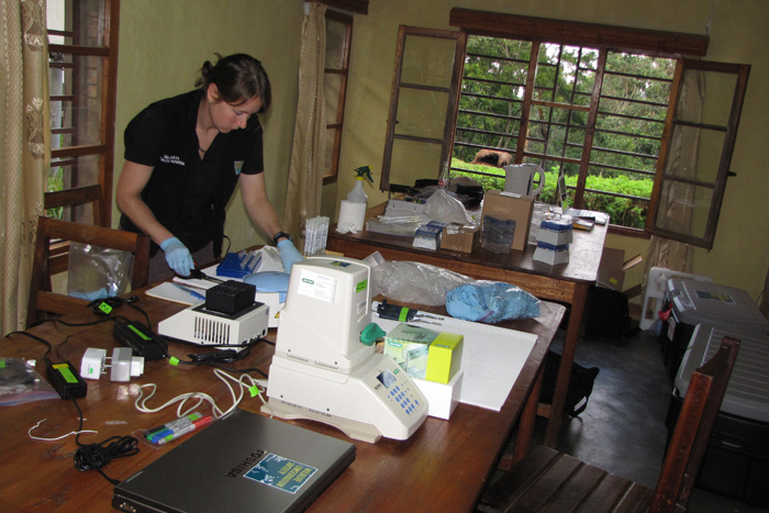
To further my interests in science and wildlife conservation, I began working in the pathology department of WCS's Zoological Health Program in 2010. Over the past five years, our department developed molecular diagnostic tools for use in zoo animals that aren't typically available in standard, commercial, veterinary diagnostic labs. We also built the mobile lab that we take to the field.
Why do we need a traveling lab? First, it often takes weeks to months, or even years, to obtain permits to export wildlife samples. Second, diagnostic expertise in wildlife infectious disease is limited in many of the remote places where we work. In the case of chytridiomycosis, at-risk populations of amphibians cannot wait. Being able to bring a laboratory to the field site eliminates that obstacle, speeding up research and analysis. The results we produce are used to document the fungus, and the mortality it causes.
The lab also provides information our team can use to educate local people and scientists about the infection, its significance, and the need for strict biosecurity measures to prevent movement of the pathogen to new areas or susceptible populations. Our hope is that, in time, our ability to provide rapid testing can be partnered with treatments for the disease as they are developed.
In addition, working on amphibian chytrid fungus in the high Andes in Peru in 2010, I took the mobile lab to Rwanda's Nyungwe Forest, a haven of biodiversity in the heart of Africa. During that trip, my colleagues and I documented the presence of chytrid fungus in the country for the first time. Luckily, infection in that case was not associated with any evidence of disease or death.
Since then, we have taken the mobile lab to Uganda, Vietnam, the Russian Far East, and twice each to Myanmar and Peru to help build in-country capacity for disease testing and to screen captive animals prior to reintroduction back into the wild.
Meanwhile, molecular technology continues to benefit from innovations and has become smaller and more portable. In fieldwork alongside glaciers in Peru last month, we field-tested a pocket-size DNA replicator that uses an iPhone as its computer and interface. Just 20 years ago, a similarly capable DNA replicator would have covered an entire tabletop. Perhaps next time you see me at the airport security checkpoint, I'll be carrying much smaller suitcases.
Follow all of the Expert Voices issues and debates — and become part of the discussion — on Facebook, Twitter and Google+. The views expressed are those of the author and do not necessarily reflect the views of the publisher. This version of the article was originally published on Live Science.
