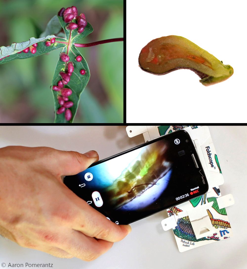
A tiny, foldable microscope that can fit in anyone's shirt pocket is revealing stunning images of nature in the jungle.
The microscope, aptly named the Foldscope, uses paper, miniscule round beads of glass, LED lights — and just a few other materials. Even crazier, the origami microscope can be placed on top of a smartphone to transmit the images it captures, so the photos can be stored and saved.
The Foldscope can magnify objects up to 2,000 times — enough to view an individual bacterium such as an E. coli. What's more, it's fairly rugged and versatile: It requires no power and can be stomped on or dropped from a three-story height without breaking, according to the Foldscope website. [See Stunning Images from the Origami Microscope]
So far, the folded-paper device is just being tested by about 10,000 early "beta testers" all around the world, but the researchers are hoping to roll out commercial development soon.
Tiny but mighty
Manu Prakash, a biophysicist at Stanford University, and his colleagues were able to develop the pocket microscope by taking advantage of the fact that a microscope is, in essence, a very simple device.
At heart, optical microscopes use a curved glass surface to focus light beams and magnify objects. To accomplish this task, the origami microscope uses tiny glass balls with different curvatures, which are placed in a tiny glass capillary tube.
Sign up for the Live Science daily newsletter now
Get the world’s most fascinating discoveries delivered straight to your inbox.
The paper body of the origami microscope can be assembled in just 10 minutes and the instructions are visual, meaning that in theory, anyone can assemble it regardless of what language he or she speaks. The materials to make a Foldscope also cost less than a dollar, meaning it could be distributed eventually to kids in the developing world, allowing them to get a closer look at nature.
Childlike rediscovery
One of the earliest users of the technology is Aaron Pomerantz, an entomologist who works with a rainforest expedition company at the Refugio Amazonas near the Tambopata Research Center in Peru.
"Some of the coolest things are from just scooping the soil up with your hands," Pomerantz told Live Science. "If you just stop and look at a tiny soil sample, it's just crawling with stuff that you have no idea what it is."
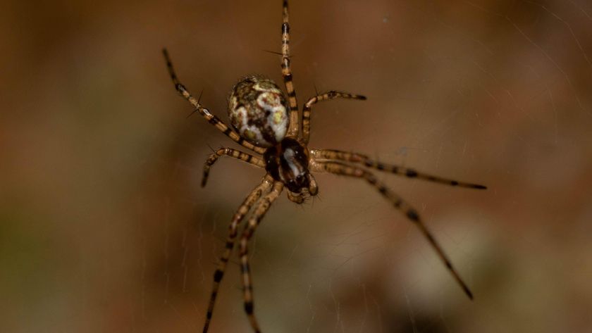
Pomerantz has taken the pocket-size device into the jungle on his nature walks, and has been amazed at the hidden beauty lurking around him in the Amazon jungle. So far, he has found a species of mite he was able to identify thanks to the microscopic images, and he used the Foldscope to pinpoint the parasite that infected an Amazon spider.
The Foldscope also revealed the larval insect responsible for bizarre, bumpy, bulblike structures on a plant known as leaf galls. Leaf galls form when insects burrow into leaves to lay eggs, which spurs leaves to release their own growth hormones, forming a makeshift home for the larva.
From teensy, alienlike insects crawling in the sand to the gorgeous pebbly texture on a flower petal, the experience has changed how Pomerantz looks at things when he goes exploring, he said.
"You say, 'Oh, I bet I can look at that under a microscope, I bet that will look weird," Pomerantz said.
Additional images from the microscope can be found at the site Microcosmos.
Follow Tia Ghose on Twitter and Google+. Follow Live Science @livescience, Facebook & Google+. Original article on Live Science.

Tia is the managing editor and was previously a senior writer for Live Science. Her work has appeared in Scientific American, Wired.com and other outlets. She holds a master's degree in bioengineering from the University of Washington, a graduate certificate in science writing from UC Santa Cruz and a bachelor's degree in mechanical engineering from the University of Texas at Austin. Tia was part of a team at the Milwaukee Journal Sentinel that published the Empty Cradles series on preterm births, which won multiple awards, including the 2012 Casey Medal for Meritorious Journalism.
