In Photos: Bee Eyes and Meat-Eating Plants Light Up Micro-Photo Contest
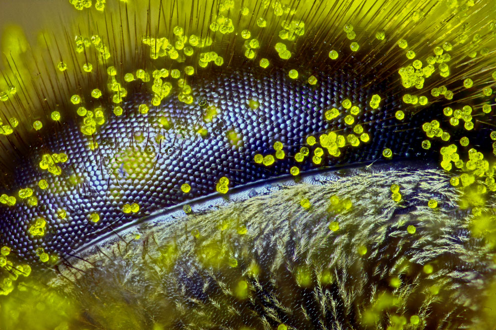
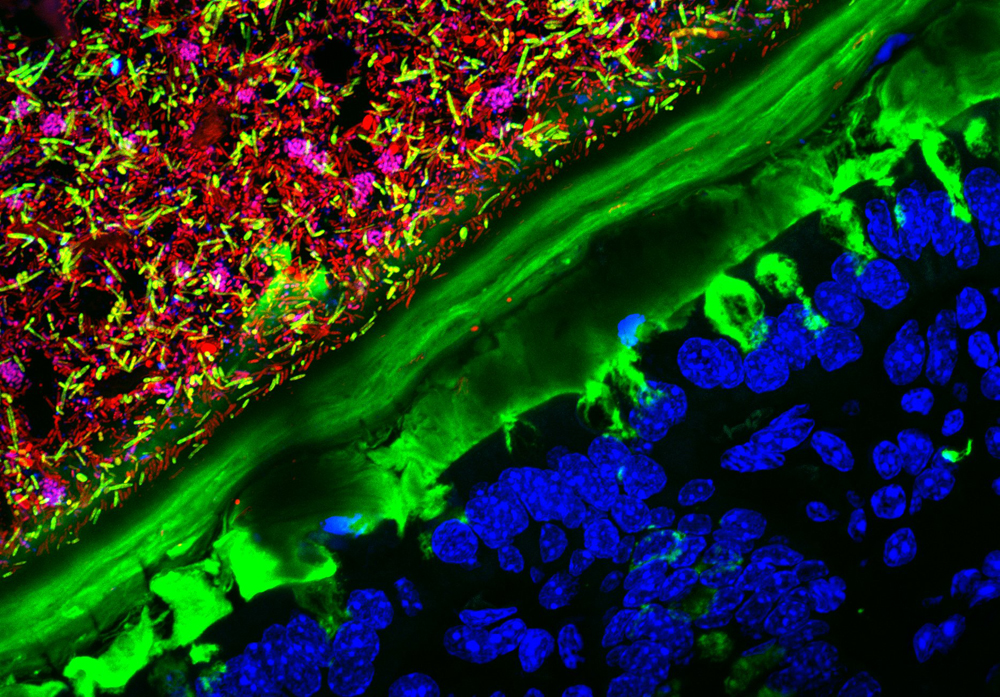
A bee's eye covered in pollen from a dandelion, a colony of bacteria growing inside a mouse's intestine, a close-up of a carnivorous plant's deadly trap — these are just a few of the winning images from this year's Nikon Small World Photomicrography competition. The annual contest offers microscope users from around the world a chance to show off their incredible photos of life's tiniest wonders. Here are this year's winning images: [Read the full story about the Small World photo contest]
1st Place
Ralph Claus Grimm
Jimboomba, Queensland, Australia
Eye of a honey bee (Apis mellifera) covered in dandelion pollen (120x), Reflected Light
2nd Place
Kristen Earle, Gabriel Billings, KC Huang & Justin Sonnenburg
Sign up for the Live Science daily newsletter now
Get the world’s most fascinating discoveries delivered straight to your inbox.
Stanford University School of Medicine, Department of Microbiology and Immunology, Stanford, California, USA
Mouse colon colonized with human microbiota (63x), Confocal
3rd Place
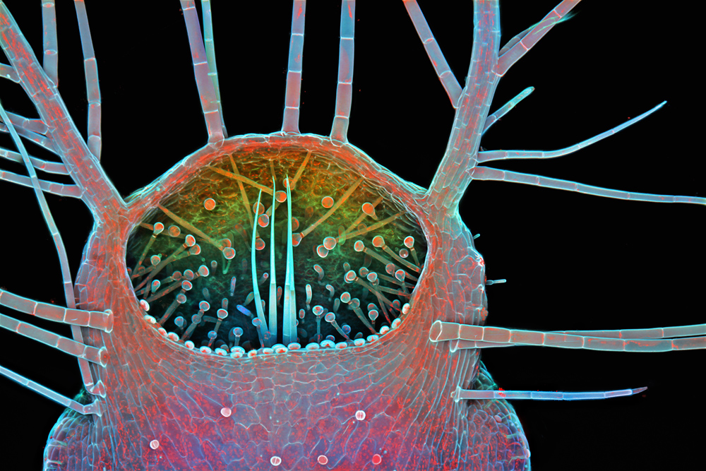
Dr. Igor Siwanowicz
Hughes Medical Institute (HHMI), Janelia Farm Research Campus, Leonardo Lab
Intake of a humped bladderwort (Utricularia gibba), a freshwater carnivorous plant (100x), Confocal
4th Place
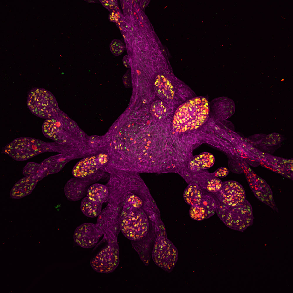
Daniel H. Miller & Ethan S. Sokol
Whitehead Institute for Biomedical Research, Massachusetts Institute of Technology, Department of Biology, Cambridge, Massachusetts, USA
Lab-grown human mammary gland organoid (100x), Confocal
5th Place
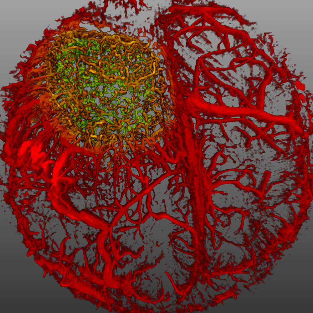
Dr. Giorgio Seano & Dr. Rakesh K. Jain
Harvard Medical School, Massachusetts General Hospital, Edwin L. Steele Laboratory for Tumor Biology, Boston, Massachusetts, USA
Live imaging of perfused vasculature in a mouse brain with glioblastoma , Optical Frequency Domain Imaging System
6th Place
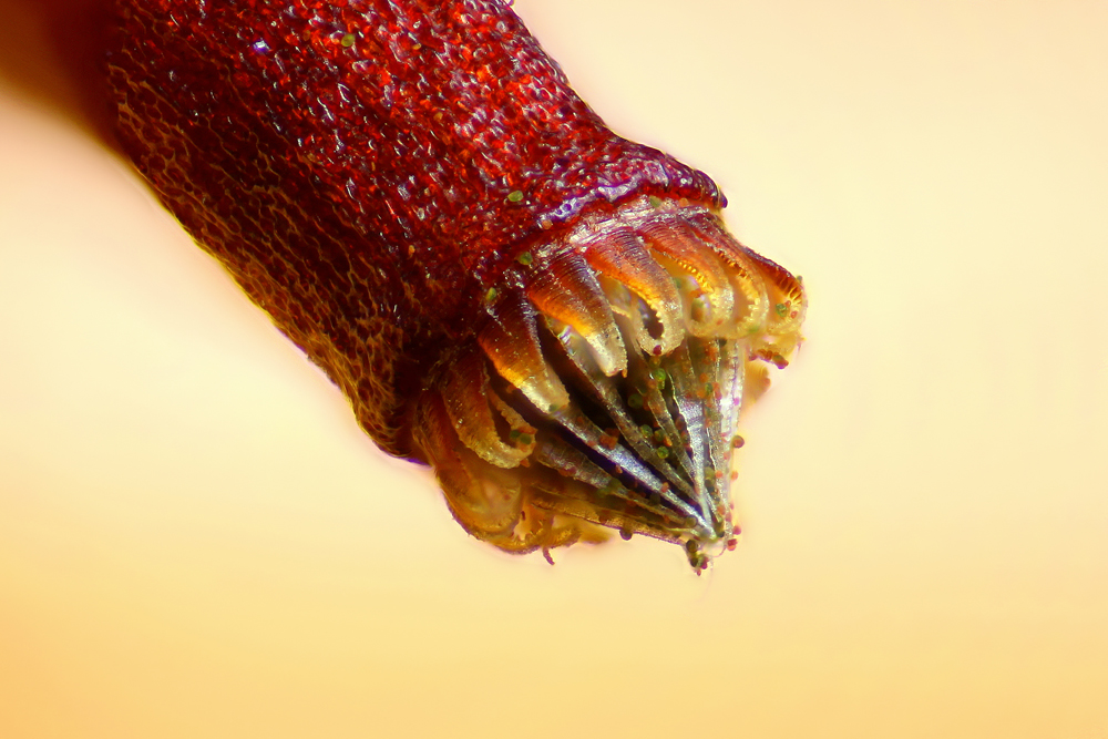
Henri Koskinen
Helsinki, Finland
Spore capsule of a moss (Bryum sp.), Reflected Light
7th Place
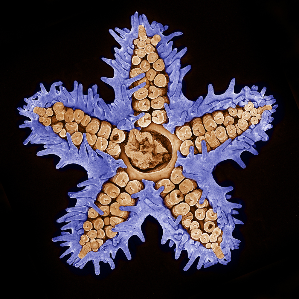
Evan Darling
Memorial Sloan Kettering Cancer Center, New York, New York, USA
Starfish imaged using confocal microscopy (10x), Confocal
8th Place
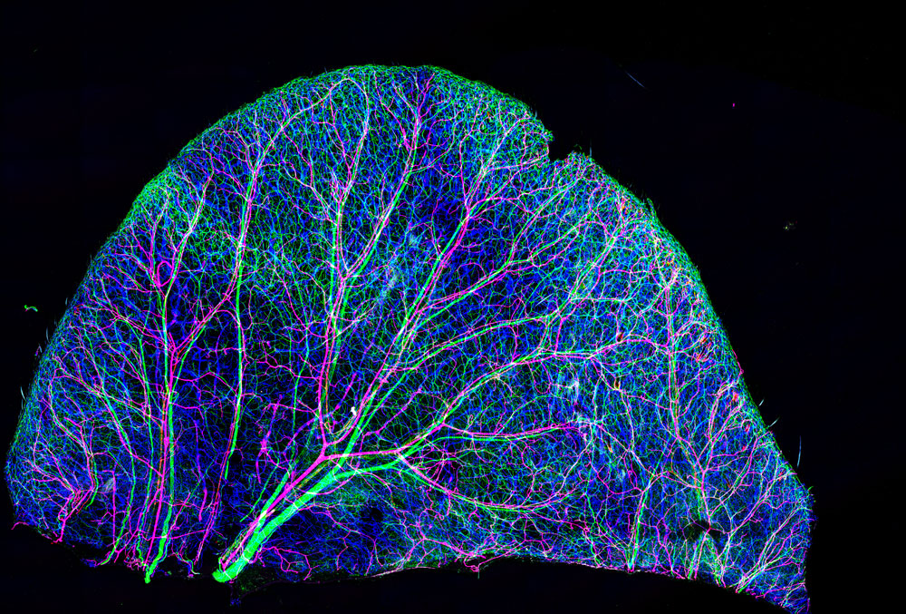
Dr. Tomoko Yamazaki
National Institutes of Health (NIH), Bethesda, Maryland, USA
Nerves and blood vessels in a mouse ear skin (10x), Confocal
9th Place
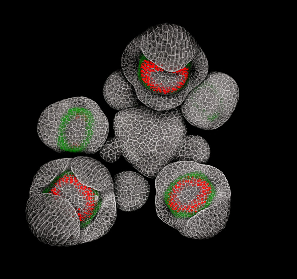
Dr. Nathanael Prunet
California Institute of Technology and Dartmouth College, Department of Biology, Pasadena, California, USA
Young buds of Arabidopsis (a flowering plant) (40x), Confocal
10th Place
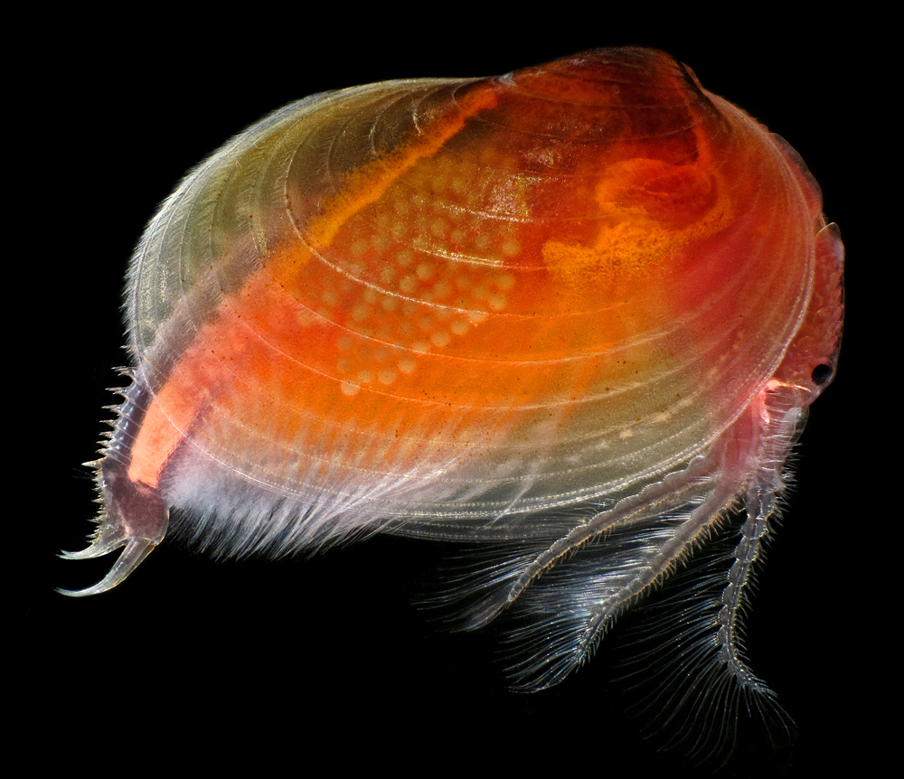
Ian Gardiner
Calgary, Alberta, Canada
Clam shrimp (Cyzicus mexicanus), live specimen (25x), Darkfield, Focus Stacking
11th Place
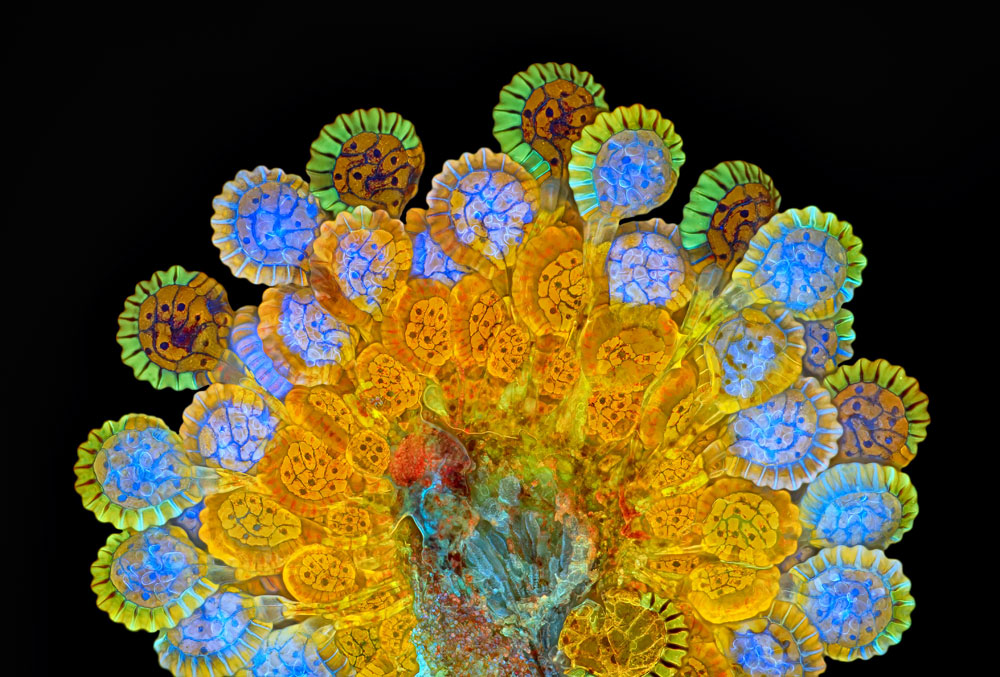
Rogelio Moreno Gill
Panama, Panama
Fern sorus at varying levels of maturity (20x), Fluorescence, Image Stacking
12th Place
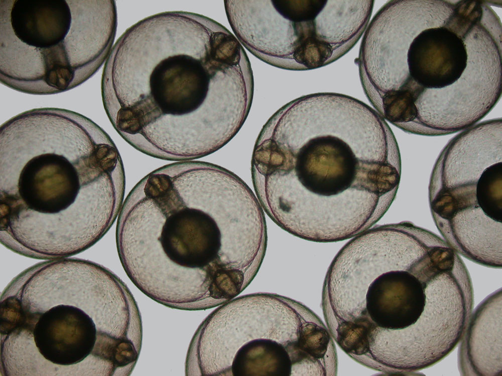
Hannah Sheppard-Brennand
Southern Cross University, National Marine Science Centre, Sydney, New South Wales, Australia
Developing sea mullet (Mugil cephalus) embryos (40x), Brightfield
13th Place
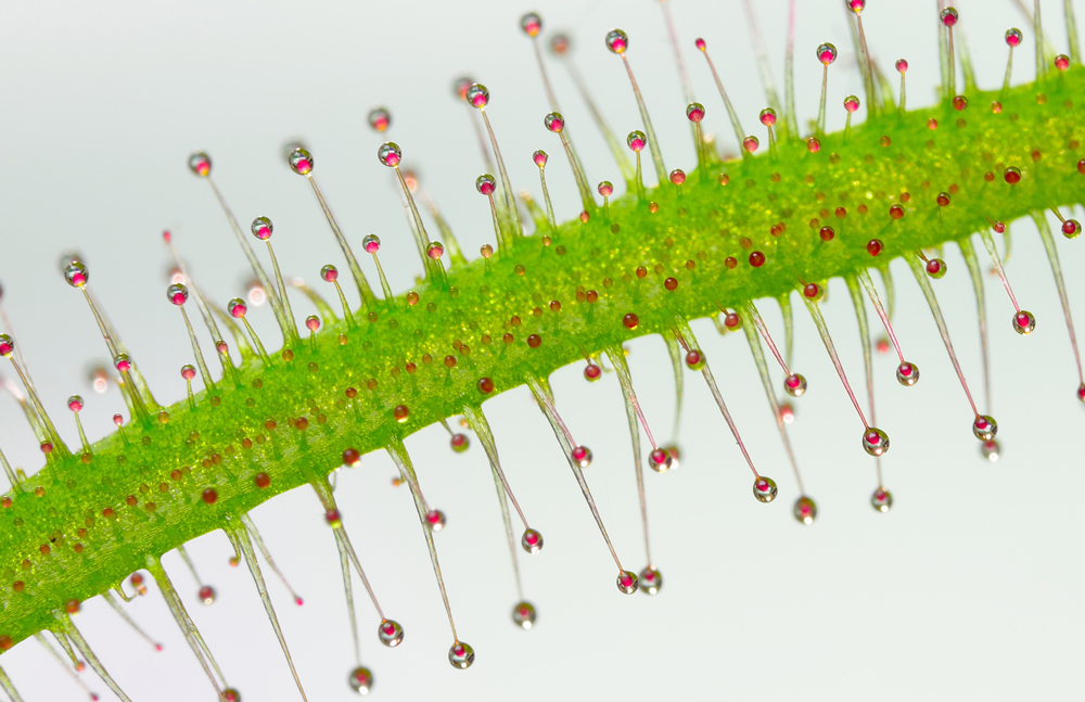
Jose Almodovar
University of Puerto Rico (UPR), Mayaguez Campus, Biology Department, Mayaguez, Puerto Rico, USA
Tentacles of a carnivorous plant (Drosera sp.) (20x), Image Stacking
14th Place
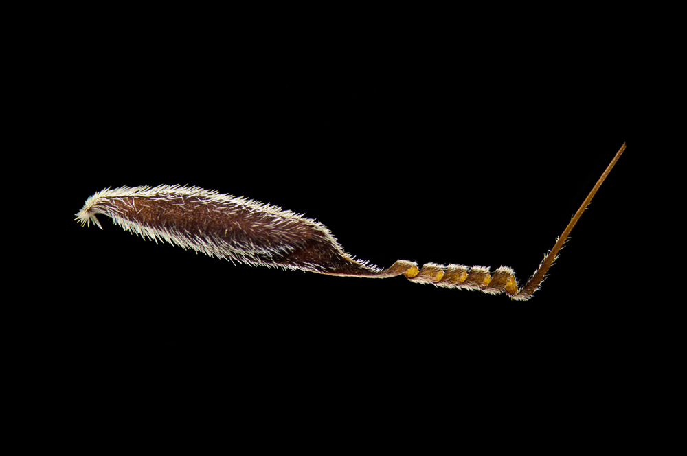
Viktor Sykora
Charles University, First Faculty of Medicine, Prague, Czech Republic
Australian grass (Austrostipa nodosa) seed (5x), Darkfield
15th Place
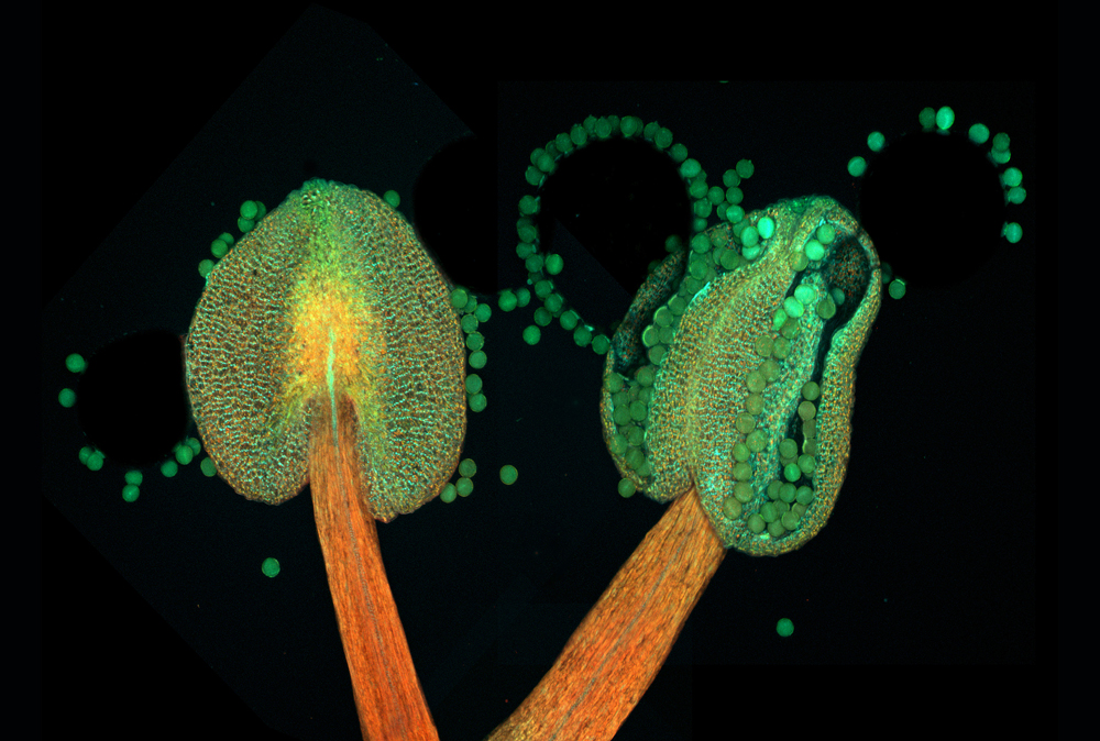
Dr. Heiti Paves
Tallinn University of Technology, Department of Gene Technology, Tallinn, Estonia
Anther of a flowering plant (Arabidopsis thaliana) (20x), Confocal
16th Place
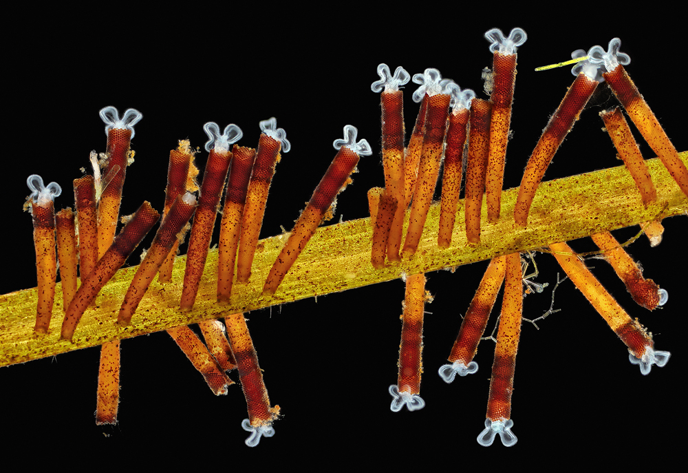
Charles B. Krebs
Charles Krebs Photography, Issaquah, Washington, USA
Feeding rotifers (Floscularia ringens) (50x), Darkfield
17th Place
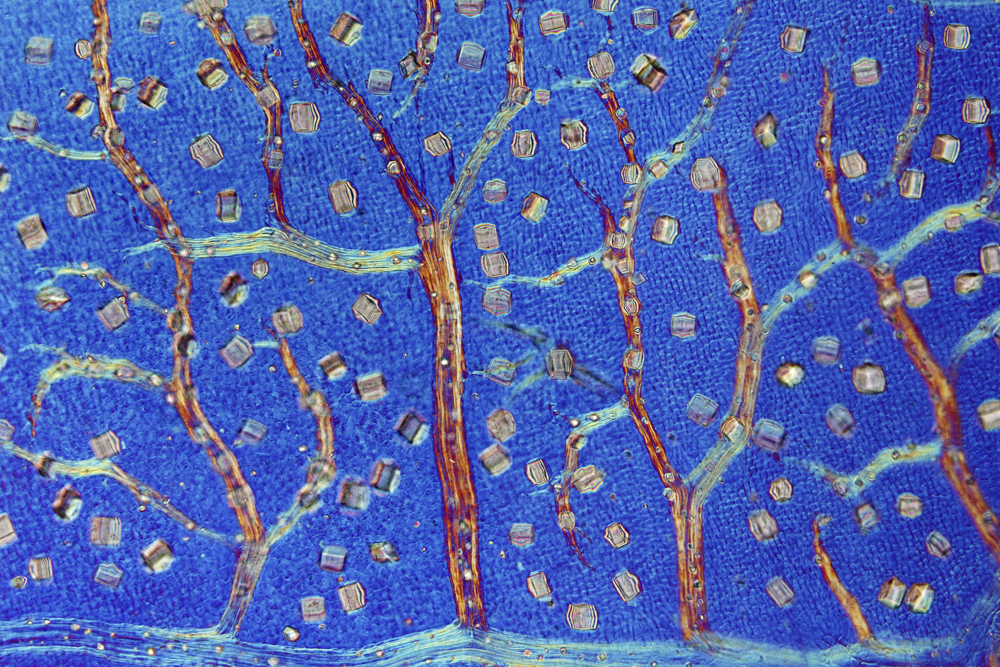
Dr. David Maitland
Feltwell, United Kingdom
Black witch-hazel (Trichodactylus crinitus) leaf producing crystals to defend against herbivores (100x), Differential Interference Contrast
18th Place
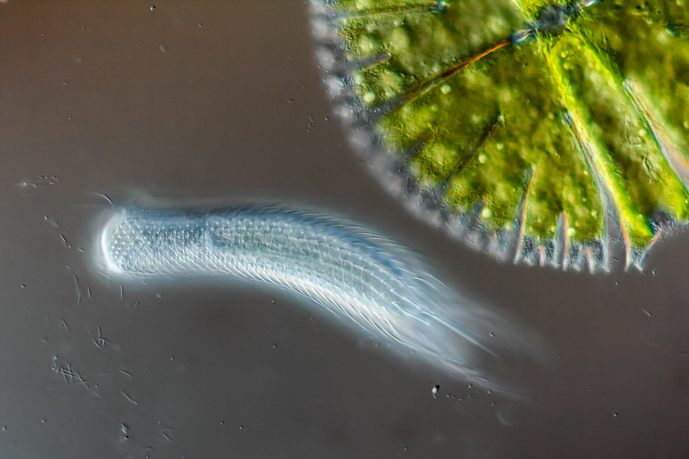
Roland Gross
Gruenen, Bern, Switzerland
Hairyback worm (Chaetonotus sp.) and algae (Micrasterias sp.) (400x), Differential Interference Contrast
19th Place
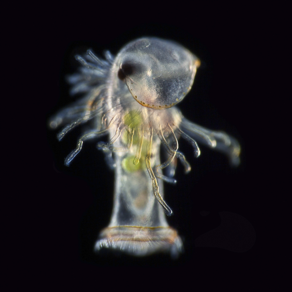
Dr. Richard R. Kirby
Marine Biological Association, Plymouth, United Kingdom
Planktonic larva of a horseshoe worm (phoronid) (450x), Darkfield
20th Place
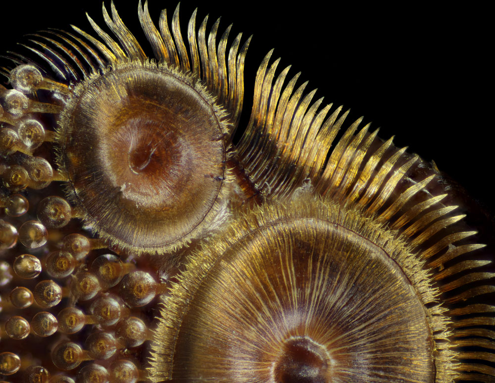
Frank Reiser
Nassau Community College, Department of Biology, Garden City, New York, USA
Suction cups on the diving beetle (Dytiscus sp.) foreleg (50x), Image Stacking, Photomerge
Baby mouse
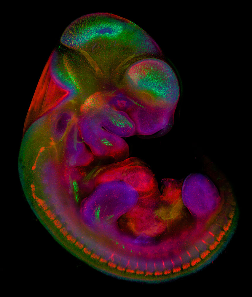
Jace Artichoker
Rochester Institute of Technology (RIT), Rochester, New York, USA
Mouse embryo, 10.5 days old, Confocal (11x)
Coral fossils
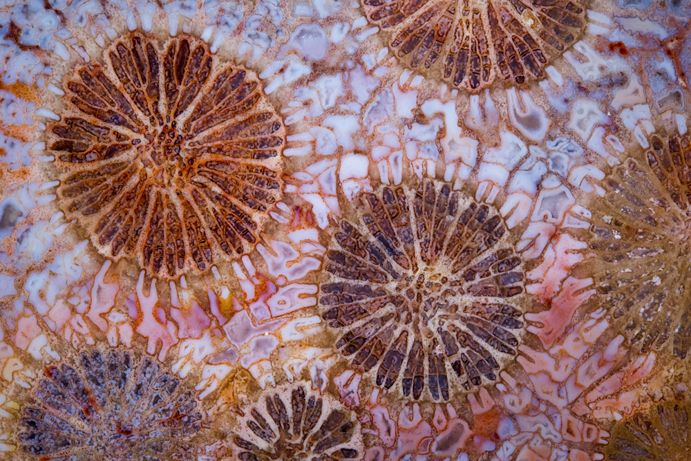
Norm Barker
Johns Hopkins School of Medicine, Department of Pathology, Baltimore, Maryland, USA
Red fossil coral slab, Reflected Light (20x)
Baby peanut worm
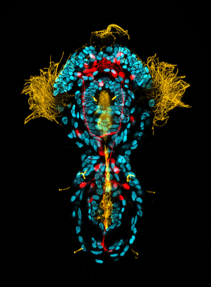
Dr. Michael J. Boyle
Smithsonian Marine Station, Life Histories Department, Fort Pierce, Florida, USA
Peanut worm (Sipuncula) trochophore larva, 3 days old (yellow: cilia; cyan: DNA; red: serotonin in the nervous system), Confocal (40x)
Adult marine worm
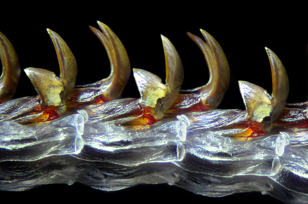
Michael Crutchley
Pembrokeshire, Wales, United Kingdom
Adult marine worm (Autolytus), Macroscopy (30x)
Cancer cell
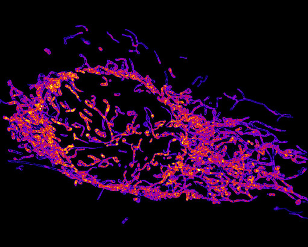
Dr. Reto Paul Fiolka
UT Southwestern Medical Center, Department of Cell Biology, Dallas, Texas, USA
Mitochondria in a live HeLa cancer cell, 3D Structured Illumination Microscopy (63x)
Stem cells
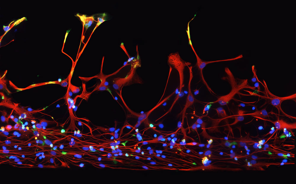
Cynthia Levinthal
Q Therapeutics, Clinical/Research Department, Salt Lake City, Utah, USA
Human neural stem cells, Fluorescence (200x)
Mouse fat
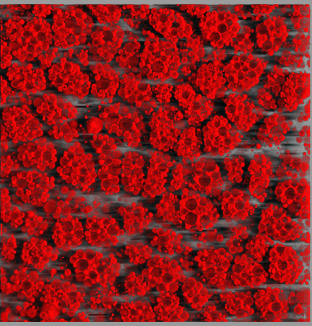
Dr. Daniela Malide
National Institutes of Health (NIH), National Heart, Lung and Blood Institute, Light Microscopy Core Facility, Bethesda, Maryland, USA
3D reconstruction of mouse brown adipose (fat) tissue, Third Harmonic Generation Microscopy (40x)
Insect bugs
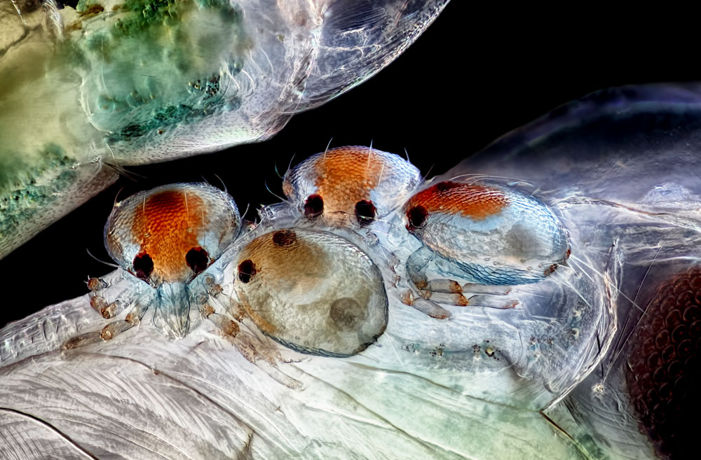
Rogelio Moreno Gill
Panama, Panama
Mites on insect pupa, Darkfield, Image Stacking (20x)
Tadpole parts
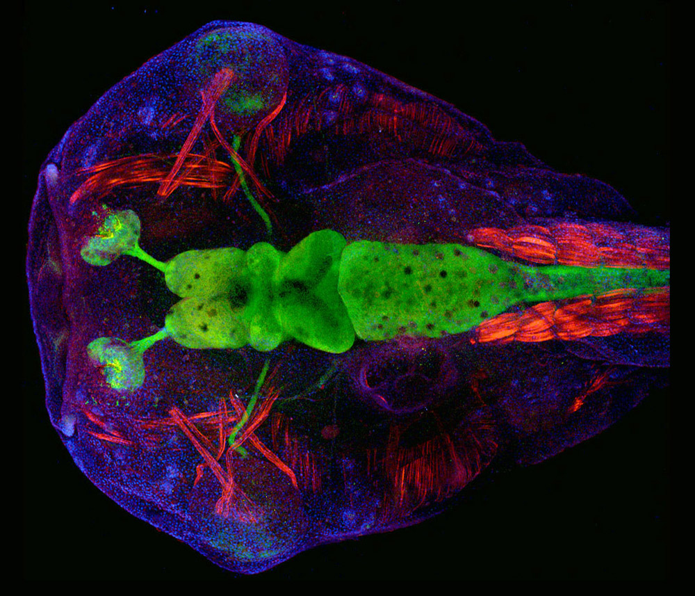
Dr. Helen Rankin
University of California, Berkeley, Berkeley, California, USA
Transgenic Xenopus laevis (African clawed toad) tadpole head expressing green neurons, Confocal (10x)
Deep sea find
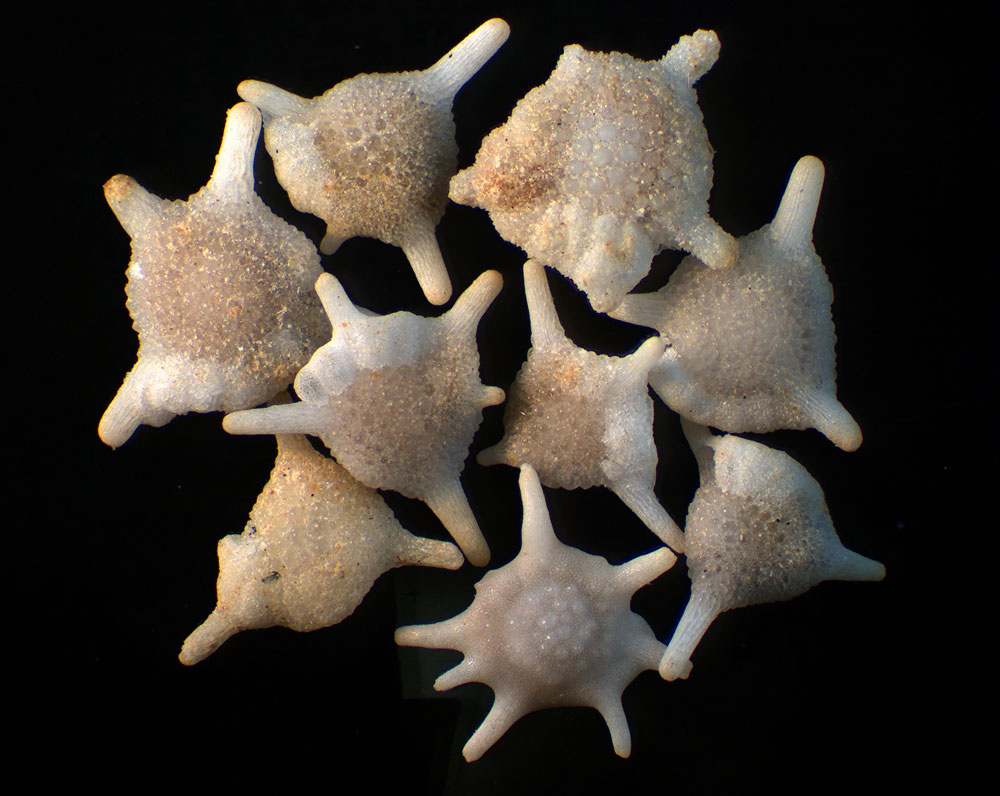
Dr. Robert B. Simmons
Briarwillow LLC, Atlanta, Georgia, USA
Foraminifera (a deep sea microscopic organism) isolated from a deep sea dredge in the Southwestern Pacific Ocean, Stereomicroscopy (4x)
Beetle bits
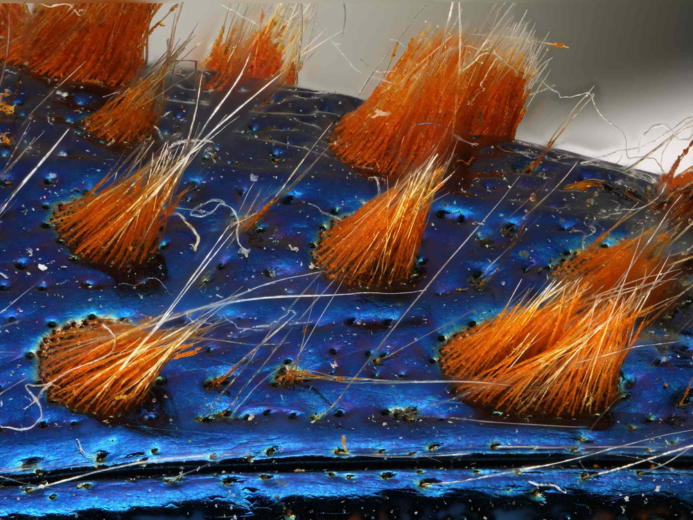
Dr. Luca Toledano
Museo Civico di Storia Naturale di Verona, Verona, Italy
Detail of jewel beetle (Coleoptera Buprestidae), Macroscopy, Image Stacking (32x)
Plant bugs
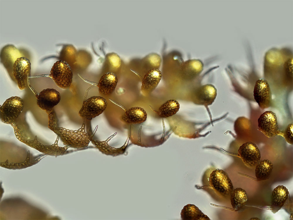
Susan Tremblay
University of California, Berkeley, Berkeley, California, USA
Liverwort (Lepidolaena taylorii) plant showing modified leaves (water sacs), which are often home to aquatic microorganisms such as rotifers, Brightfield (100x)
Colony of individuals
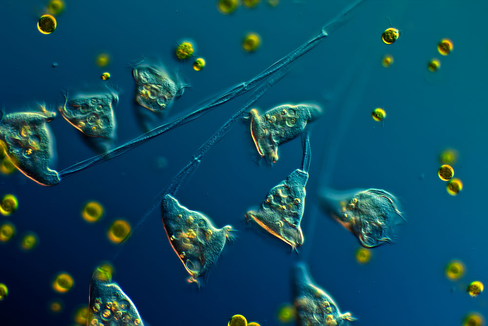
Arturo Agostino
Reggio Calabria, Italy
Colony of single celled organisms (Carchesium ciliates) (160x), Differential Interference Contrast
Glowing alga
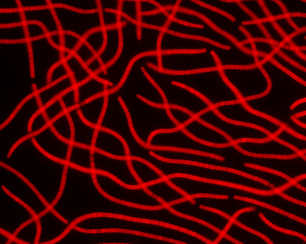
Dr. Kesara Margrét Anamthawat-Jonsson, Andrey N. Gagunashvili & Ólafur S. Andrésson
University of Iceland, Institute of Life and Environmental Sciences, Reykjavik, Iceland
Nostoc, a blue-green alga (cyanobacteria) showing red autofluorescence of the chlorophylls (400x), Fluorescence
Muscles
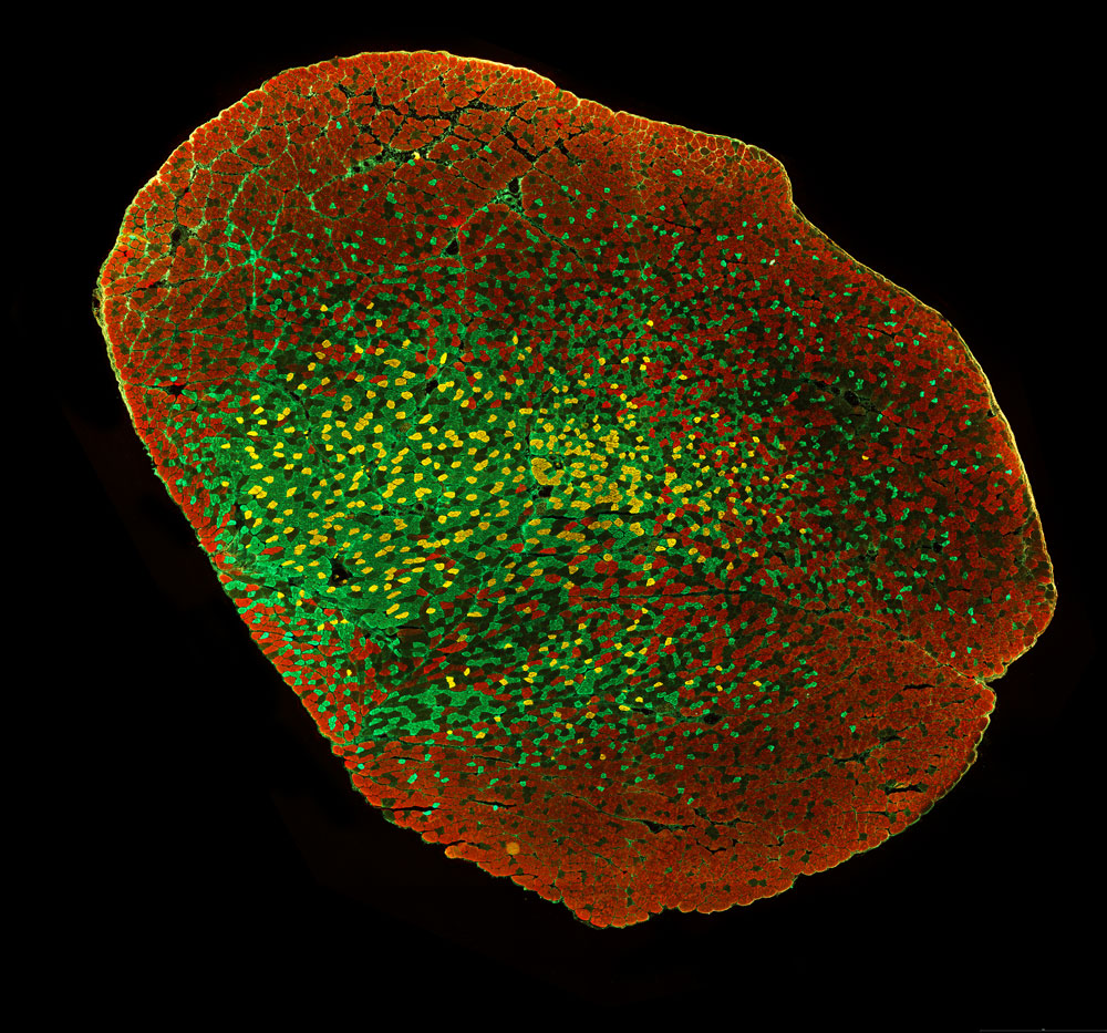
Dr. Konstantin D. Bergmeister, Marion Gröger, Martin Aman, Anna Willensdorfer, Krisztina Manzano-Szalai & Oskar C. Aszmann
Medical University of Vienna, Christian Doppler Laboratory for Restoration of Extremity Function, Division of Plastic and Reconstructive Surgery, Depa
Murine biceps muscle stained to show different muscle fiber populations (20x), Fluorescence
Oh the colors
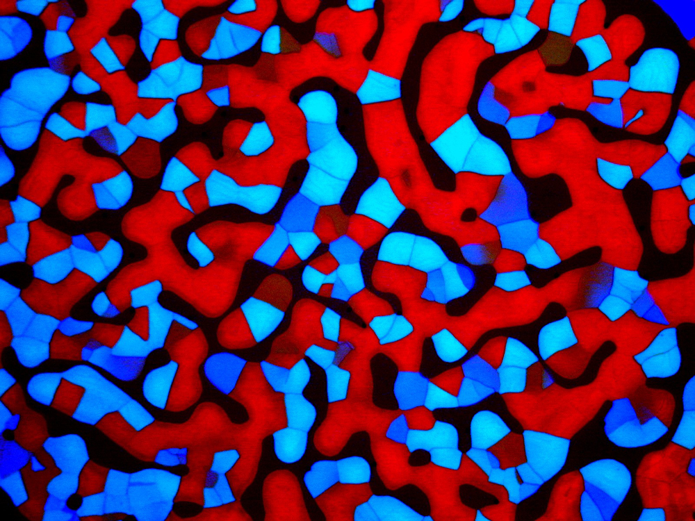
Christian Bohley
Martin Luther University Halle-Wittenberg, Halle (Saale), Germany
Degenerating Blue Phases (II) of 55% CB15 in E48 (substance used in manufacture of Liquid Crystal Displays) (100x), Polarized Light
Fruit fly larva
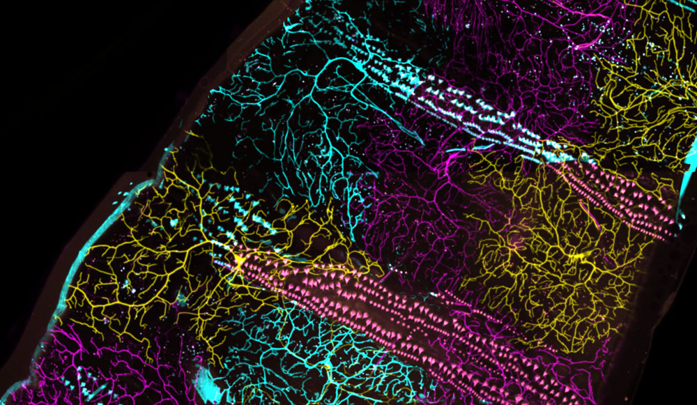
Dr. Maria Boulina, Dr. Akira Chiba & Hasitha Samarajeewa
University of Miami, Miami, Florida, USA
Individually colored neurons in a live fruit fly (Drosophila) larva, Fluorescence, Confocal
Tiny numbers
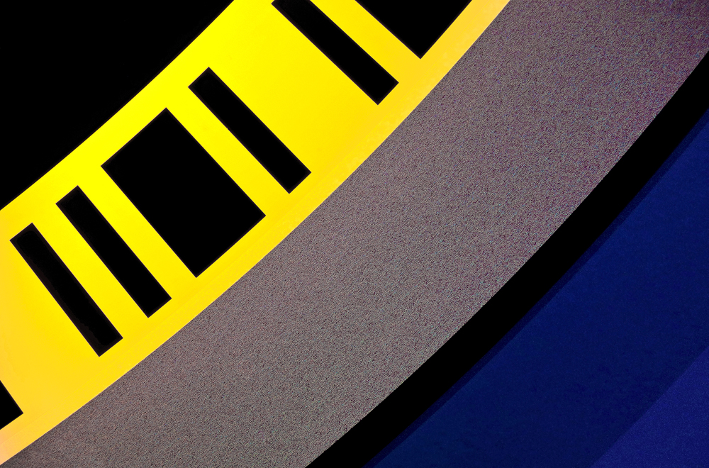
Massimo Brizzi
Empoli, Italy
Numerical traces on a DVD/Blu-ray (100x), Fiber Optic Illumination
Mouse brains
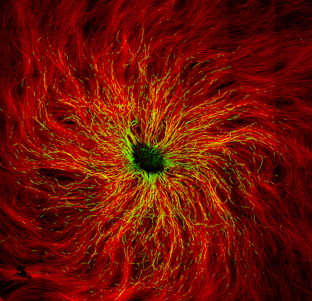
Dr. Alessio Colombo
DZNE, Munich, Bavaria, Germany
Mouse dorsal root ganglia (neuronal plus Schwann cells) in culture (10x), Confocal
Dinner time
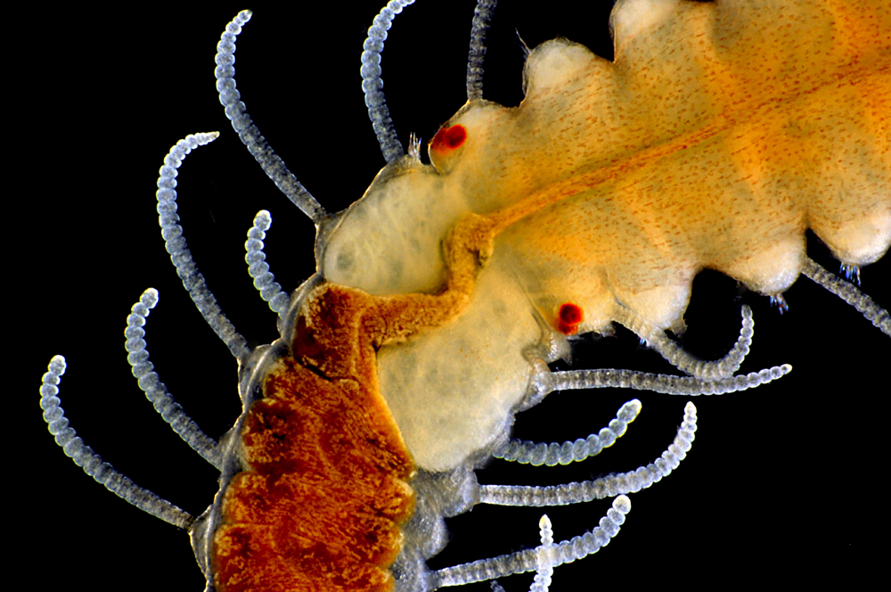
Michael Crutchley
Pembrokeshire, Wales, United Kingdom
Radula (feeding structure) of an aquatic snail (Limpet) (40x), Darkfield Epi.
Rat brains
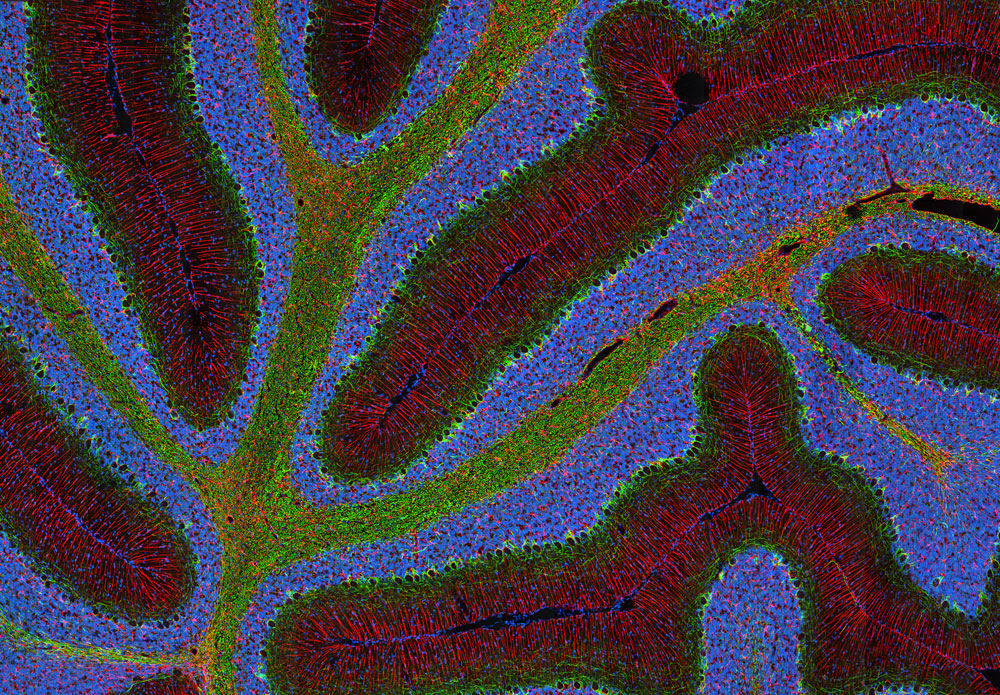
Thomas Deerinck
University of California, San Diego, La Jolla, California, USA
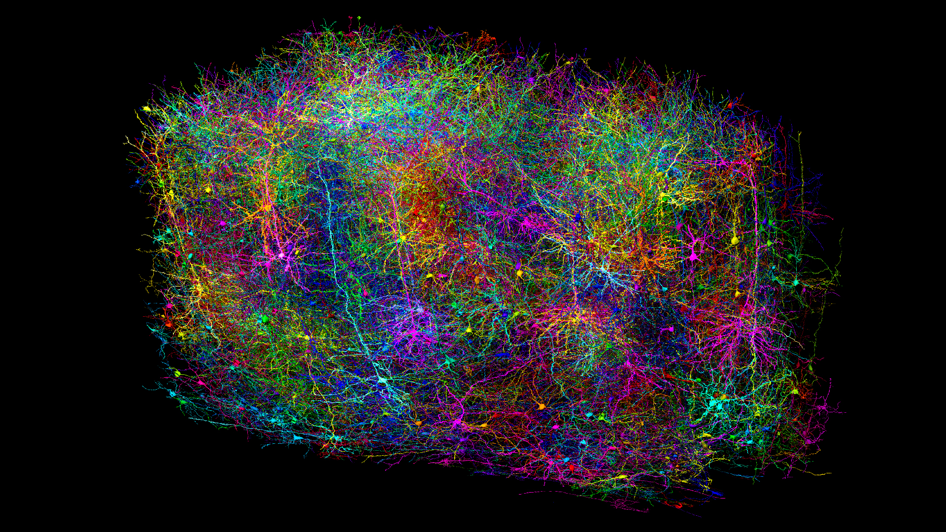
Triple-labeled rat cerebellum (100x), Multiphoton
Mouse eye
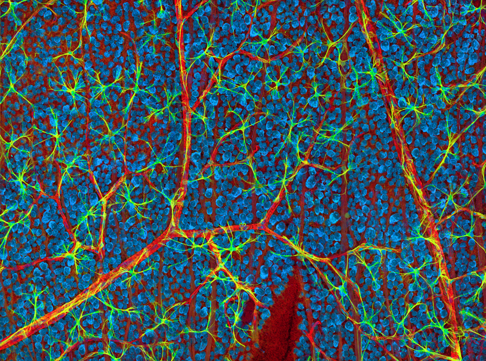
Thomas Deerinck
University of California, San Diego, La Jolla, California, USA
Vasculature and glial cells in the optic fiber layer of a mouse retina (200x), Confocal
Spider parts
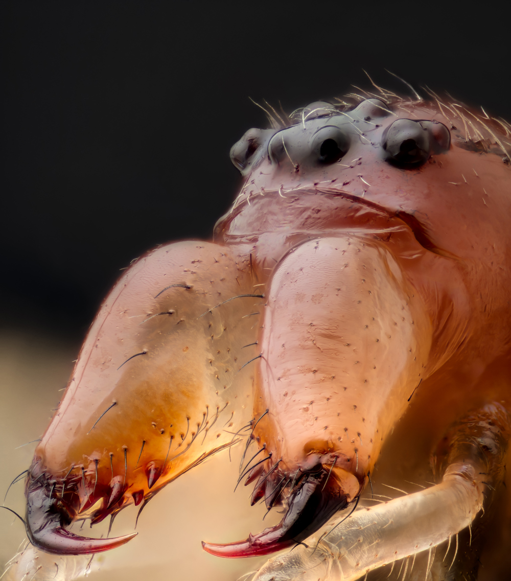
Geir Drange
Asker, Norway
Jaws and head of a long jawed spider (Metellina sp.) (10x), Reflected Light
Fossils
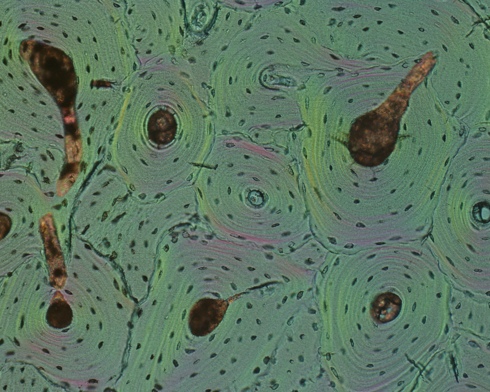
Dr. Santiago Gomez
University of Cadiz, Cadiz, Spain
Hipparion fossilized bone (100x), Polarized Light
Peace lily pollen
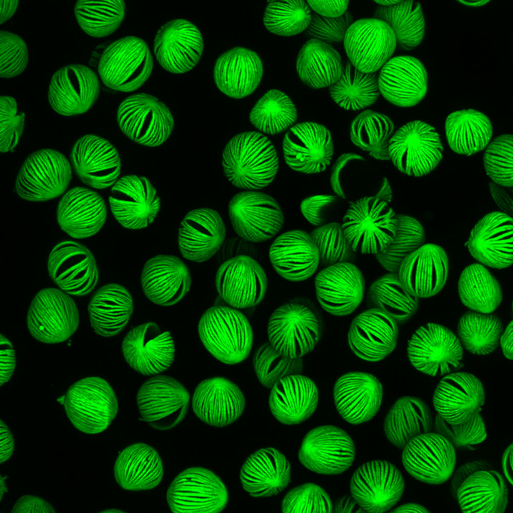
Dr. Marta Guervos
University of Oviedo, University Institute of Oncology of Asturias, Scientific-Technical Facilities/Optical Microscopy Unit, Oviedo, Asturias, Spain
Pollen grains of a peace lily (Spathiphyllum sp.) (63x), Confocal
Fabric and glue
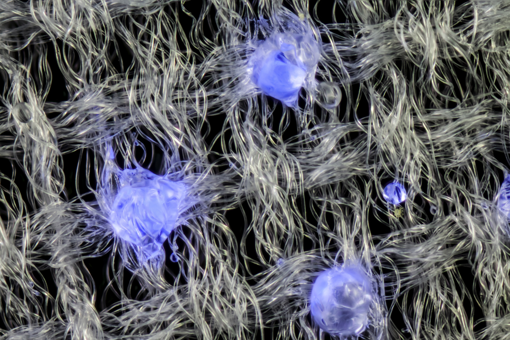
Gerd-A. Günther
Düsseldorf, Germany
Vilene fabric (glue drops shown in blue) (80x), Brightfield, Fluorescence
Crystal
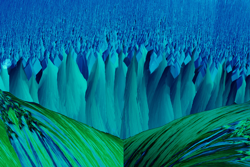
Dr. John Hart
University of Colorado Boulder, Department of Atmospheric and Oceanic Sciences, Boulder, Colorado, USA
Resorcinal and methylene blue crystal (33x), Polarized Light
Sea urchin skin
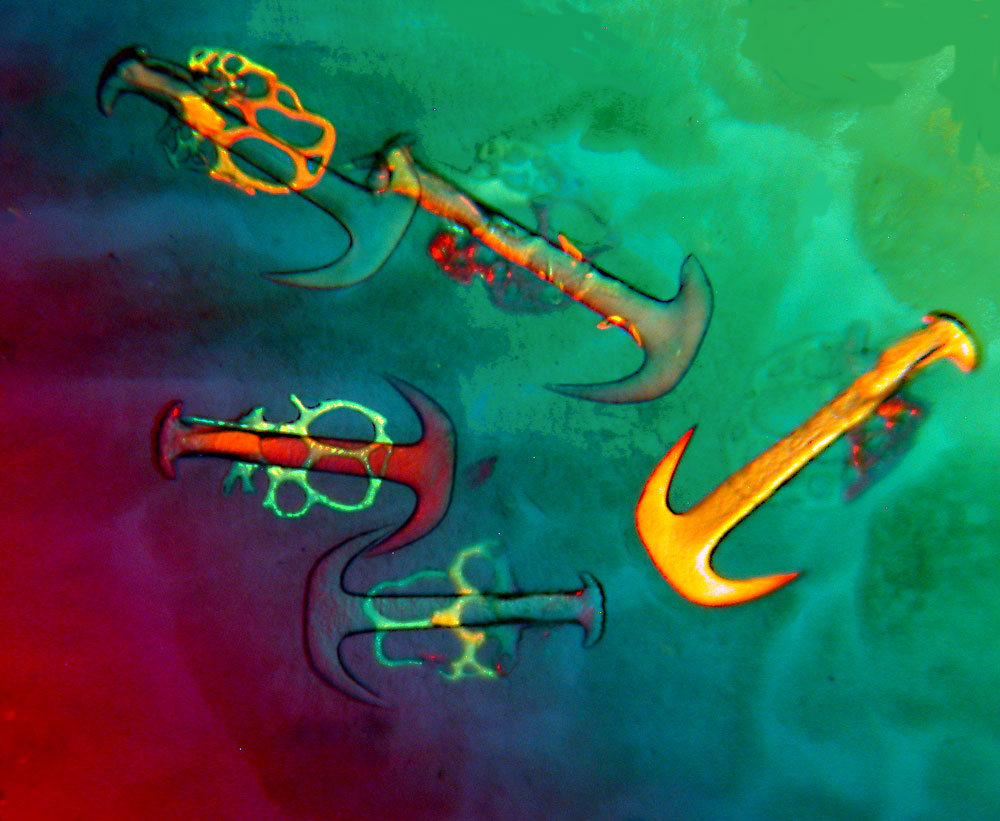
Dr. Richard Howey
University of Wyoming, Department of Philosophy, Laramie, Wyoming, USA
Skin of a sea urchin (Synapta) containing plates and anchors composed of calcareous (chalky) material (63x), Polarized Light
Mouse tongue
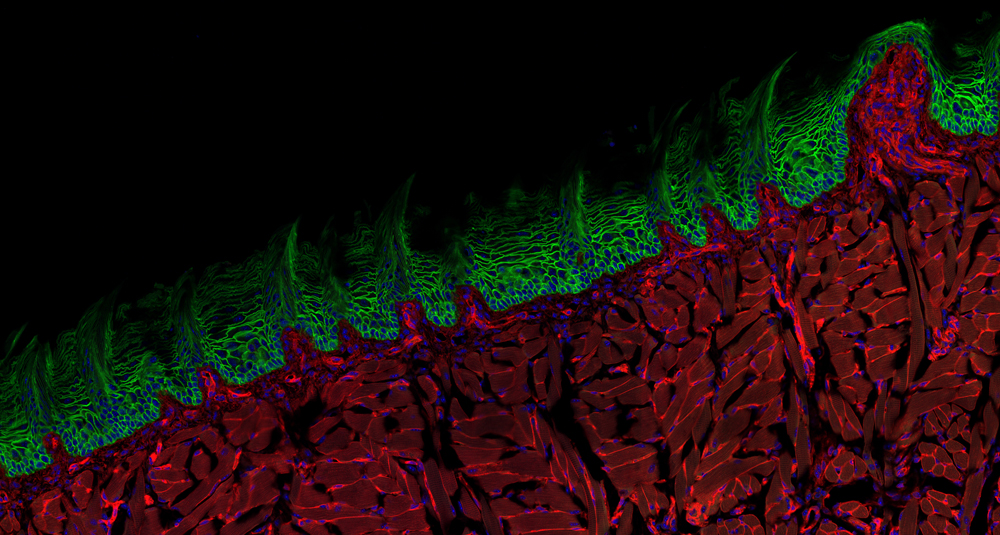
Dr. Matthew Kofron & Tayaramma Thatava
Cincinnati Children’s Hospital Medical Center, Cincinnati, Ohio, USA
Mouse tongue section (60x), Confocal
Cow lung
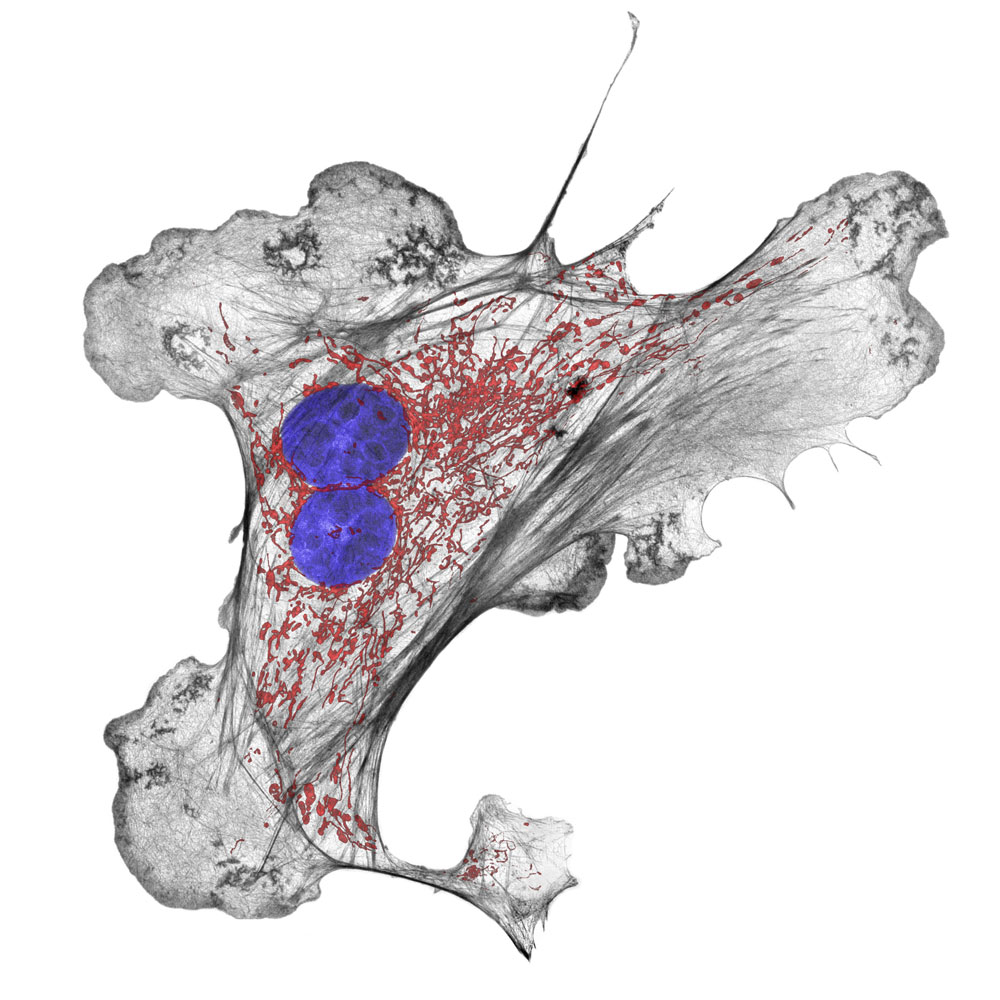
Dr. Talley J. Lambert
Harvard Medical School, Department of Cell Biology, Boston, Massachusetts, USA
Cow lung artery cell highlighting the cellular components actin (black), mitochondria (red), and DNA (blue) (60x), 3D-Structured Illumination Microscopy
Vampire moth mouth
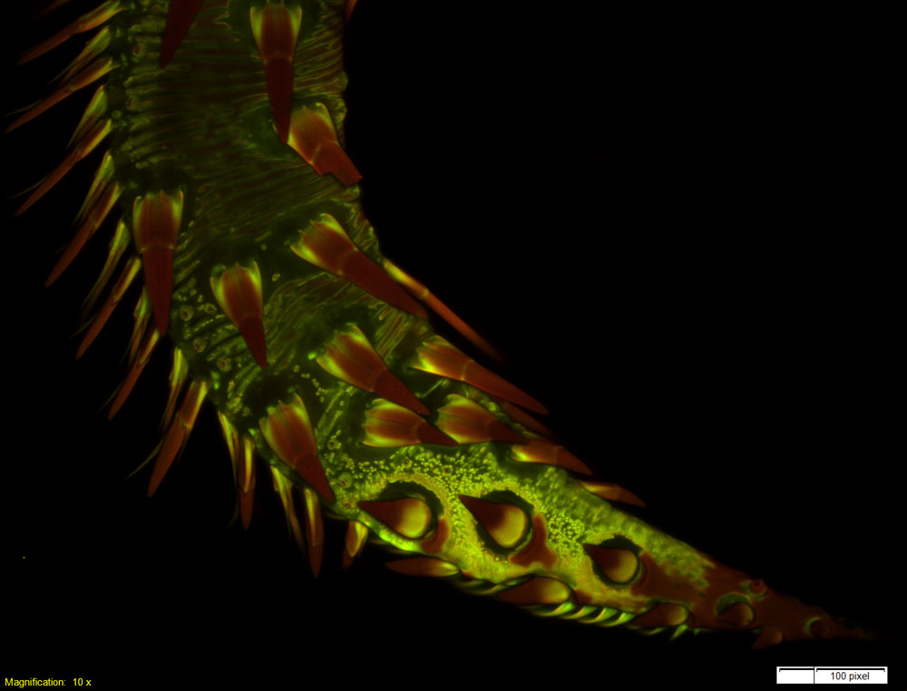
Dr. Matthew S. Lehnert
Kent State University at Stark, North Canton, Ohio, USA
Proboscis (mouthparts) tip of a vampire moth (Calyptra thalictri). The modified tip and tearing hooks (red) assist with piercing fruit and mammal tissues for feeding (10x), Confocal
Honey bee stinger
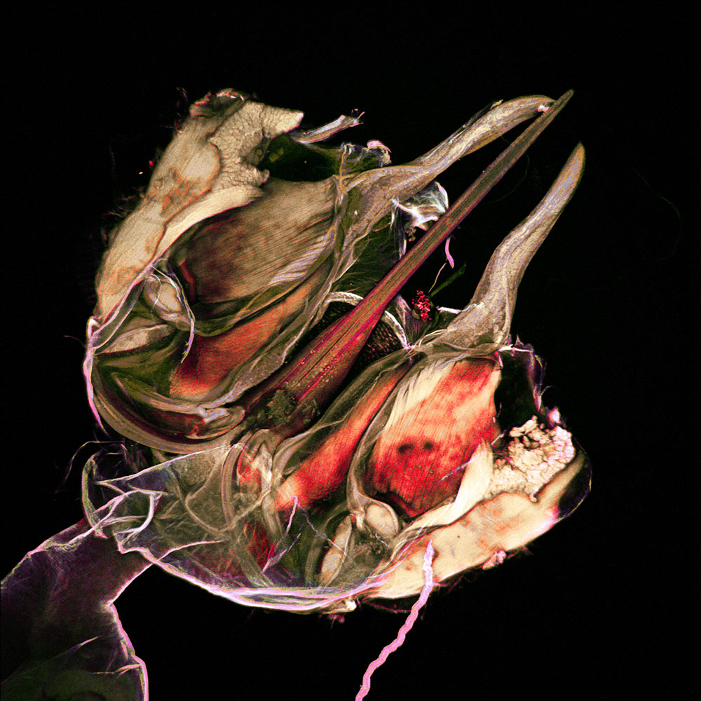
Harry Leung
Harvard Medical School, Program in Cellular and Molecular Medicine, Children's Hospital Boston, Boston, Massachusetts, USA
Stinger of a honey bee (20x), Confocal
Floating organs
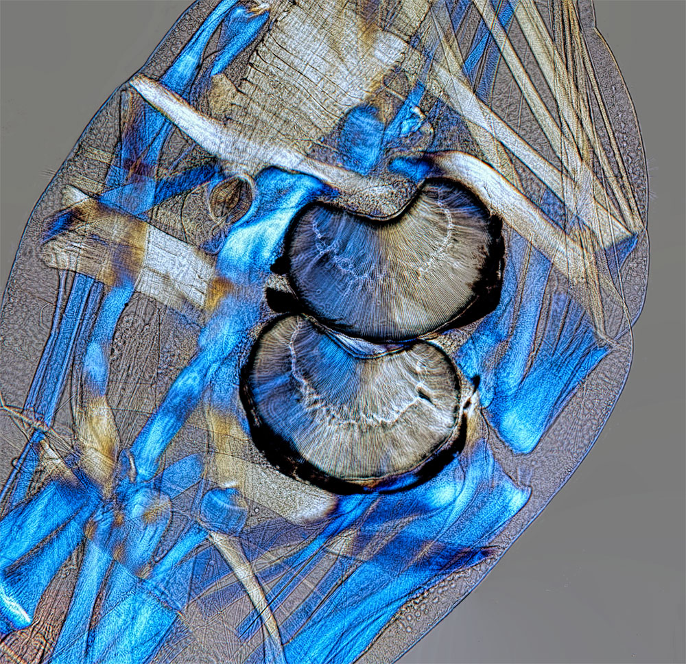
Dr. David Linstead
Bromley, Kent, United Kingdom
Buoyancy organs of a phantom midge (Chaoborus) larva (125x), Polarized Light
Pottery details
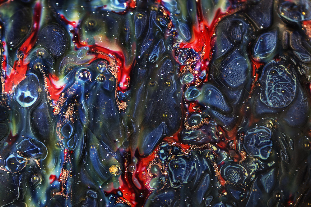
Yvonne (Yi-Chieh) Lu
New York, New York, USA
Detail of ancient Chinese pottery from the Song Dynasty (960-1126 AD) (4x), Macroscopy
Ancient engravings
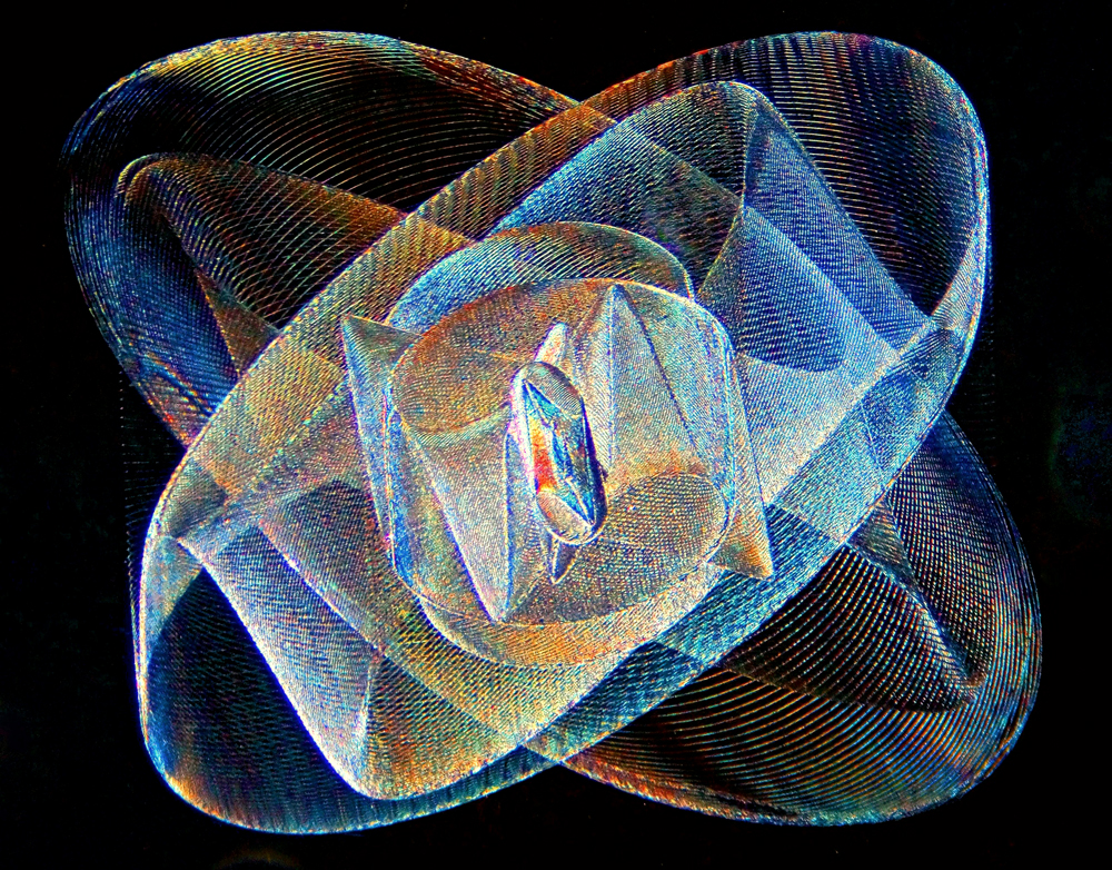
Howard Lynk
Morehead City, North Carolina, USA
Microengraving on glass from an antique microscope slide created by Washington Teasdale c. 1880 (100x), Darkfield
Water lily leaf
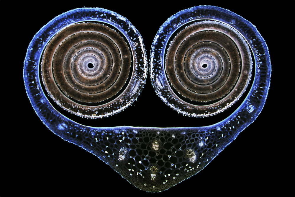
Dr. David Maitland
www.davidmaitland.com, Feltwell, United Kingdom
Leaf cross section of a water lily leaf bud (Nupha lutea) (12.5x), Brightfield
Papyrus bundles
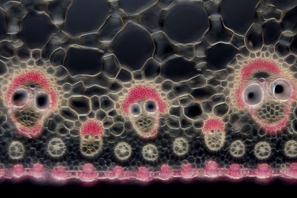
Dr. David Maitland
www.davidmaitland.com, Feltwell, United Kingdom
Vascular bundles of papyrus (Cyperus papyrus) (200x), Differential Interference Contrast
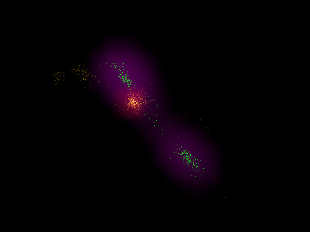
Cell division
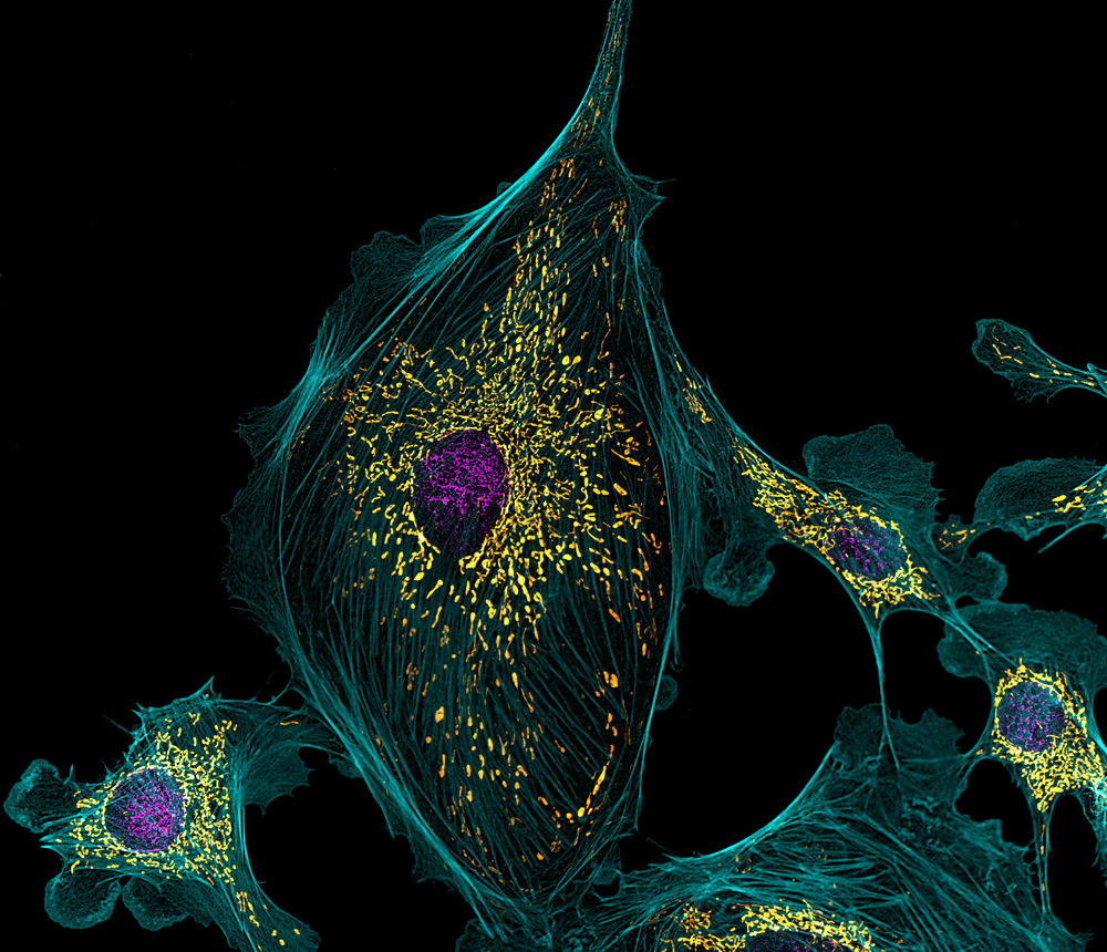
Dr. Robert Markus & Dr. Jafar Mahdavi
University of Nottingham, School of Life Sciences, Super Resolution Microscopy Department, Nottingham, United Kingdom
Bacterial DNA and peptide probe binding inside bacteria following division (DNA-YOYO-1: green pixels for high res & purple for wide field; Cy5-probe: yellow pixels for high res & orange for wide field), STORM Super-Resolution Microscopy and Widefield Fluorescence
Bovine heart
Dr. Robert Markus
University of Nottingham, School of Life Sciences, Super Resolution Microscopy Department, Nottingham, United Kingdom
Bovine pulmonary artery endothelial cells (1100x), Structured Illumination Microscopy
Electrodes
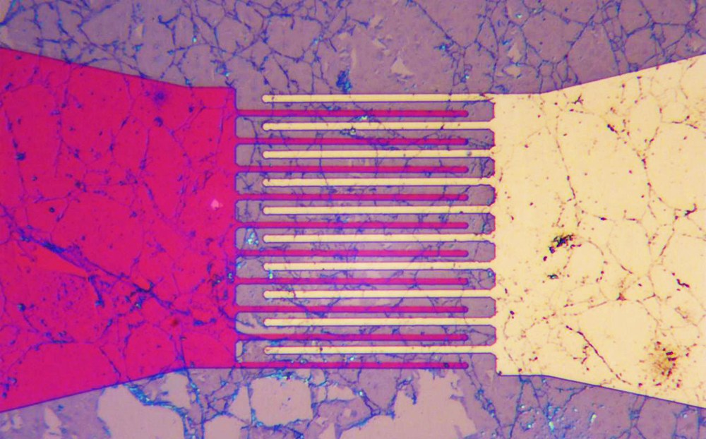
Dr. Aleksandar Matkovic
University of Belgrade, Institute of Physics, Center for Solid State Physics and New Materials, Belgrade, Serbia
Gold and titanium electrodes covered by graphene sheet (500x), Brightfield
Ostrich fern
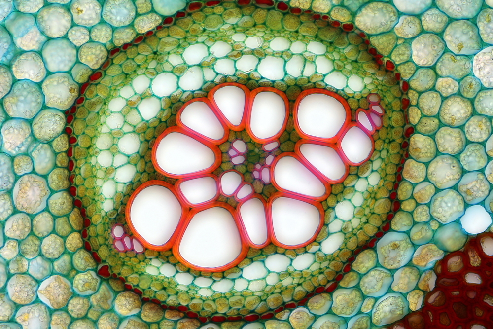
Anatoly Mikhaltsov
Omsk, Russia
Transverse section of an ostrich fern (250x), Brightfield
Mouse embryo
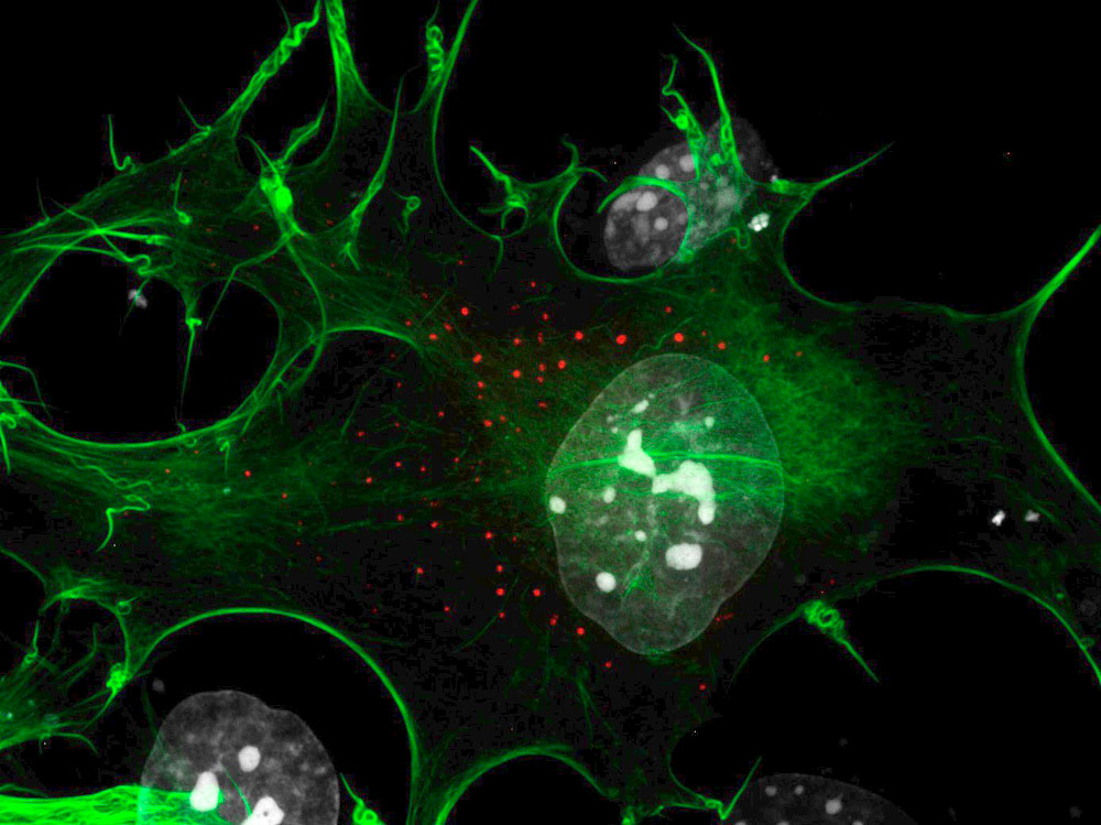
Dr. Tetsuaki Miyake
York University, Department of Biology, Toronto, Ontario, Canada
Cultured mouse embryo cells (100x), Confocal Live-Cell Imaging (Orthogonal Projection of the Z-Stack Images)
Rocks
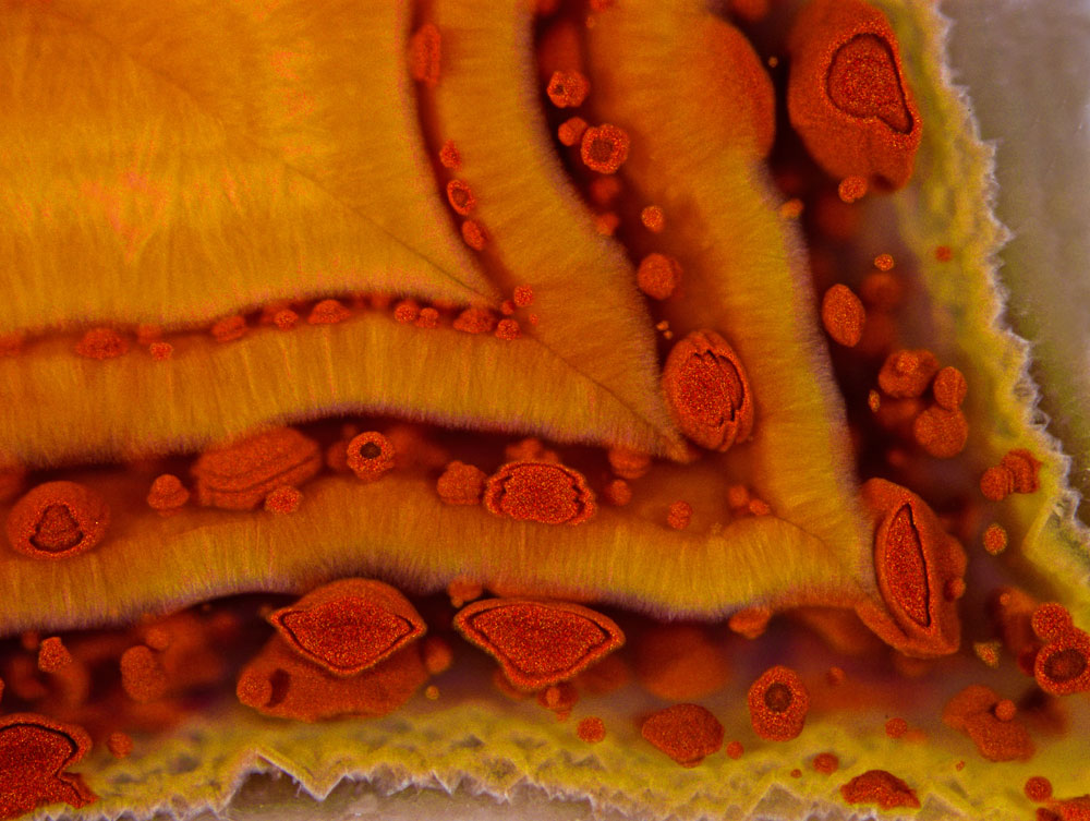
Douglas Moore
University of Wisconsin - Stevens Point, University Relations and Communications, Stevens Point, Wisconsin, USA
Fairburn agate from Black Hills of western South Dakota (63x), Fiber Optic Illumination
Tiny organism
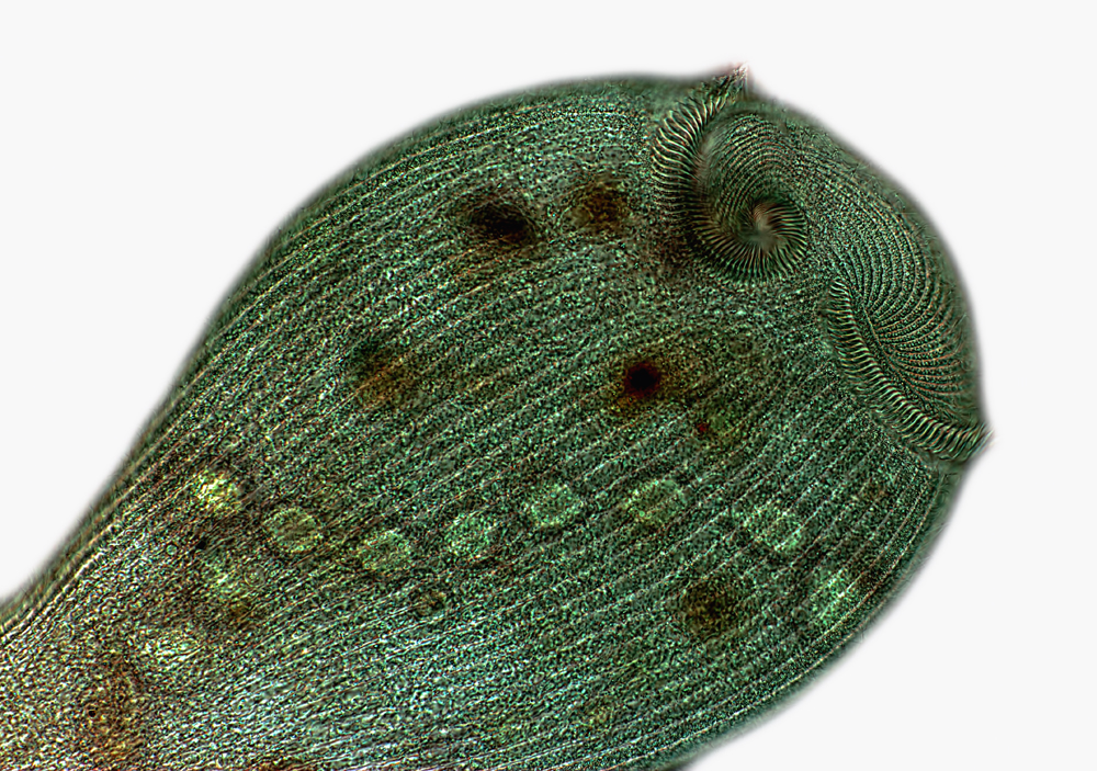
Rogelio Moreno Gill
Panama, Panama
Stentor showing the macronucleus and peristome cilia (40x), Brightfield
Water flea
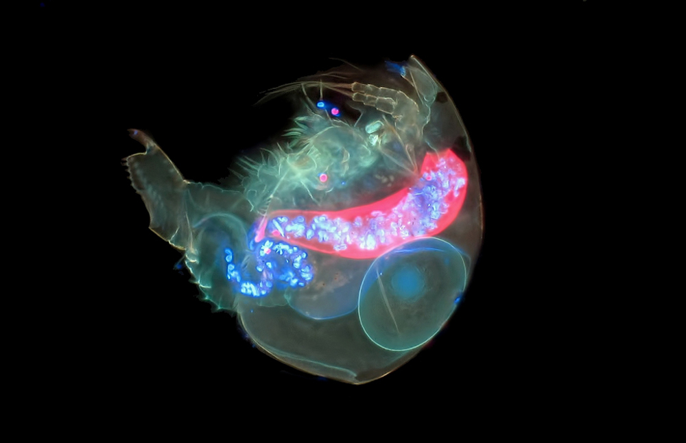
Jacek Myslowski
Wloclawek, Kujawsko-Pomorskie, Poland
Alona guttata (water flea) (200x), Fluorescence
Stressed cells
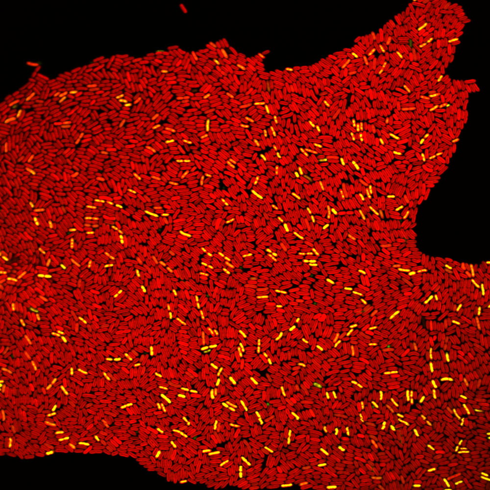
Robert Newby
Seton Hall University, Department of Biological Sciences, South Orange, New Jersey, USA
Autofluorescence and viability overlay in zinc stressed cyanobacterium Synechococcus sp. IU 625. Cells in red are healthy; yellow are impaired; and green are dead (600x), Fluorescence
Sea shells
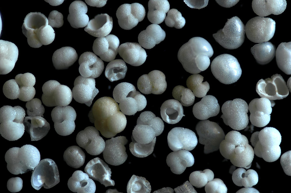
Caoimhghin Ó Maolagáin
National Institute of Water and Atmospheric Research, Wellington, New Zealand
A scatter of foraminifera shells from the sea (40x), Reflected Light
Garnet and magnetite
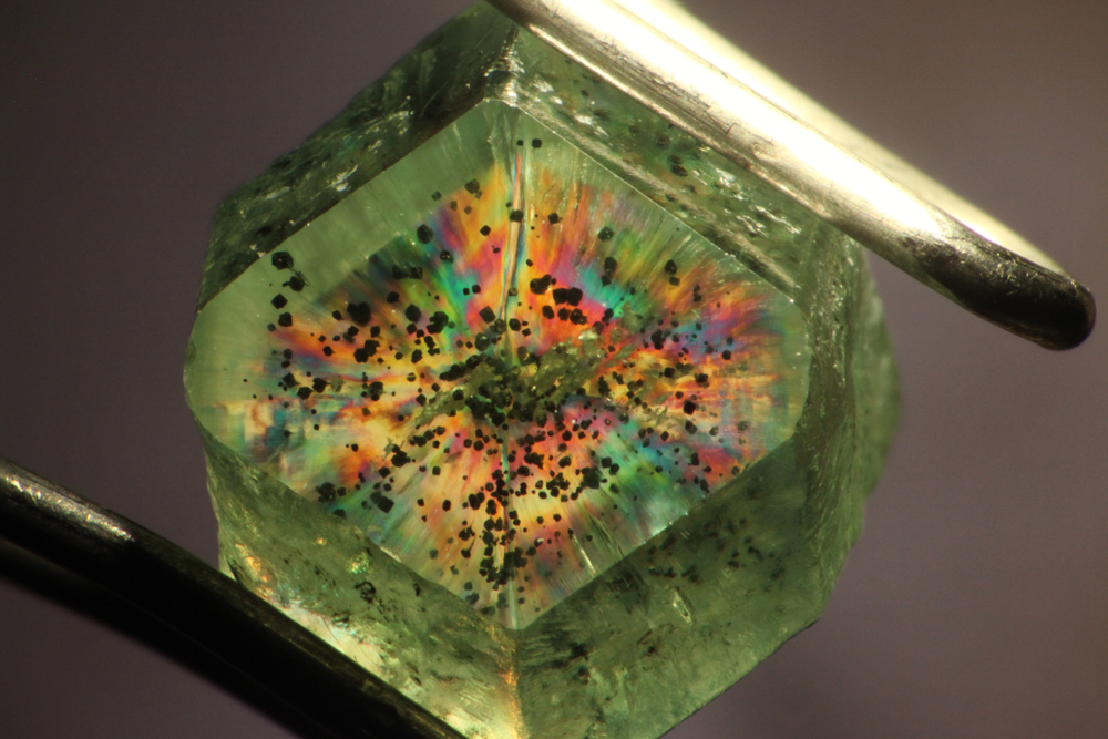
Dr. Aaron Palke
Gemological Institute of America, Carlsbad, California, USA
Birefringent andradite garnet with magnetite inclusions (15x), Polarized Light
Rove beetle head
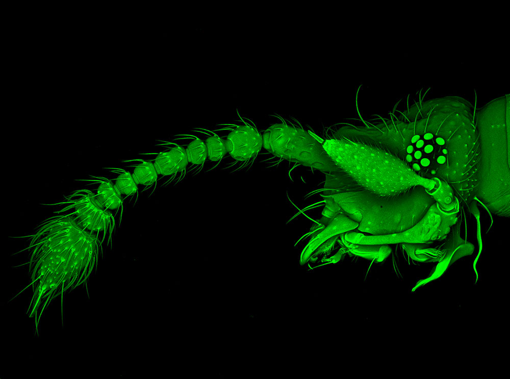
Dr. Joseph Parker
Columbia University, Department of Genetics and Development, New York, New York, USA
Rove beetle head (Tychobythinus sp.) (10x), Confocal
Moth wing scales
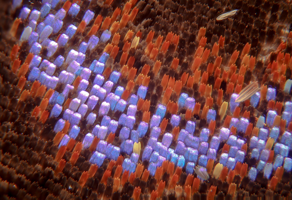
Donald Parsons
Madison, Wisconsin, USA
Scales on moth wing (300x), Image Stacking
Tadpole
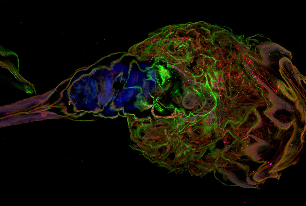
Katherine Pfister
University of Virginia, Charlottesville, Virginia, USA
Xenopus laevis (frog) tadpole- ventral view (10x), Confocal
DNA cell nucleus
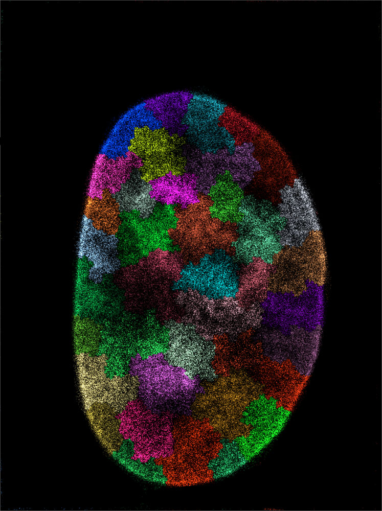
Kirti Prakash
Institute of Molecular Biology, Mainz, Mainz, Germany
DNA inside cell nucleus, Super-Resolution Microscopy
The changing
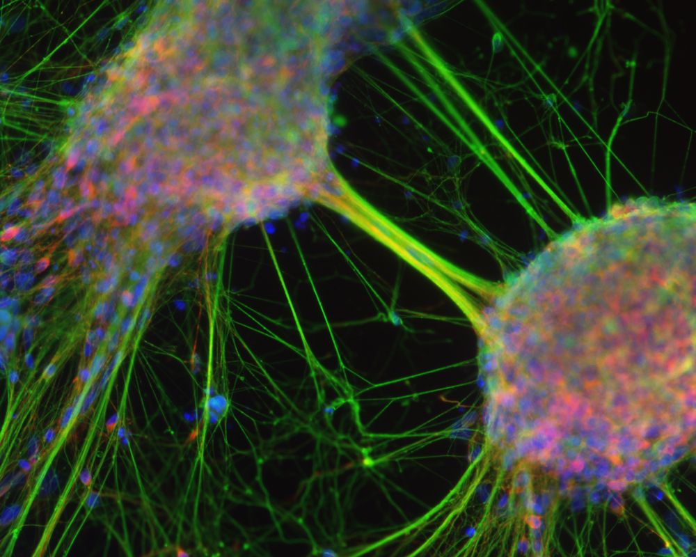
Dr. Ariadna Recasens
University of Sydney, Kolling Institute of Medical Research, Sydney, Australia
Micrometamorphosis: from human stem cells to neurons (20x), Fluorescence
Fungus mold
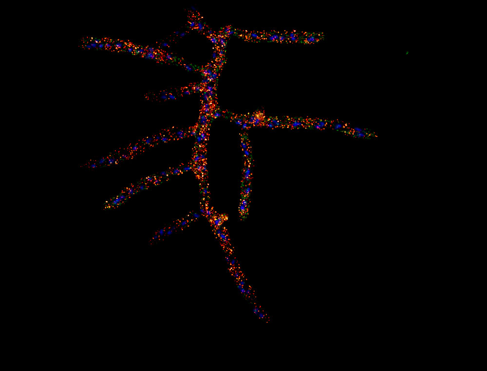
Samantha Roberts & Dr. Amy Gladfelter
Dartmouth College, Department of Biological Sciences, Hanover, New Hampshire, USA
Fungus mold (Ashbya gossypii sp.) labeled for intracellular structures (63x), Fluorescence
Photon emission
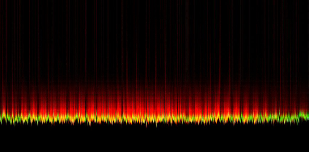
Rebecca Saleeb, Robert Henderson & Paul Dalgarno
Heriot-Watt University, Institute of Biological Chemistry, Biophysics and Bionengineering, Edinburgh, United Kingdom
The single photon emission pattern of a cell in time, Fluorescence Lifetime Imaging
Ocean trash
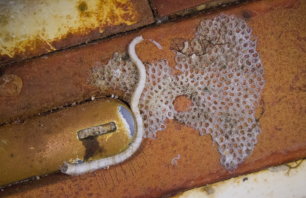
Dr. Robert B. Simmons
Briarwillow LLC, Atlanta, Georgia, USA
Power button of a cellular phone (collected from ocean bottom near Kefalonia, Greece). Features include the remains of encrusting bryozoans and a calcareous tube made by a marine worm (4x), Stereomicroscopy
Fishing net
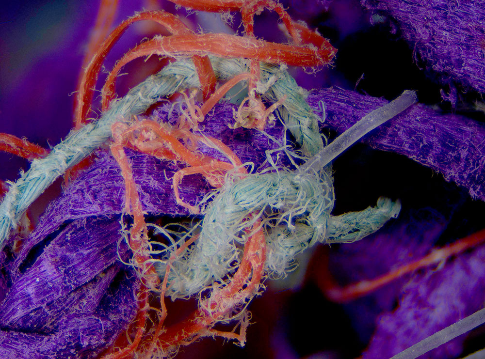
Dr. Robert B. Simmons
Briarwillow LLC, Atlanta, Georgia, USA
Plastic components of a drifting fishing net (ghost net) recovered from the ocean (5x), Stereomicroscopy
Male moth antenna
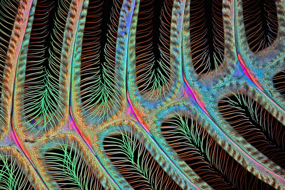
Dr. Igor Siwanowicz
Ashburn, Virginia, USA, Hughes Medical Institute (HHMI), Janelia Farm Research Campus, Leonardo Lab
Antenna of a male moth (Anisota sp.) (100x), Confocal
Blowfly mouth parts
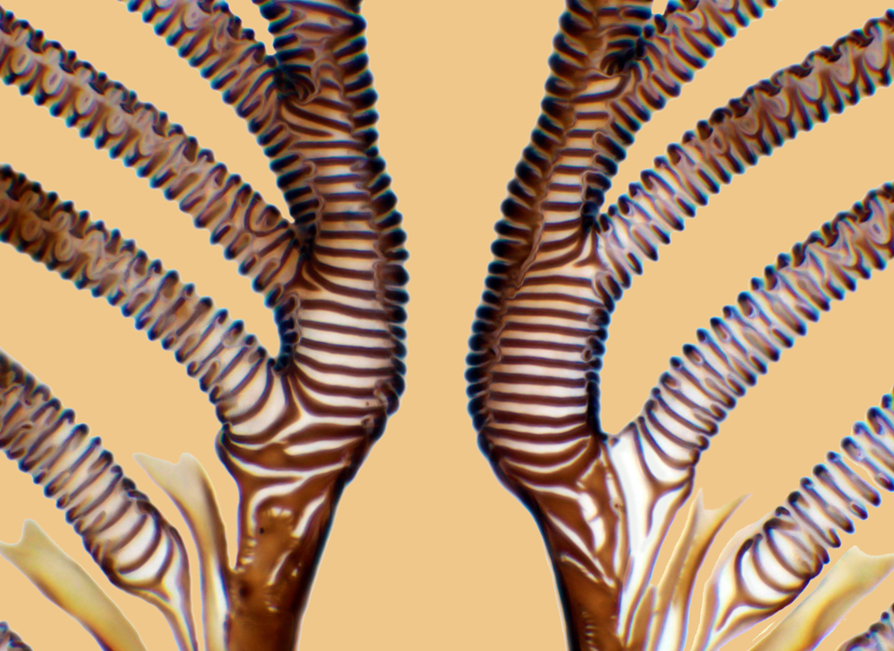
Raymond Morrison Sloss
Quekett Microscope Club, Banbury, Oxfordshire, United Kingdom
Mouth parts (pseudo trachea) of a Blowfly (Calliphora vomitoria) (750x), Brightfield
Dicot plant root
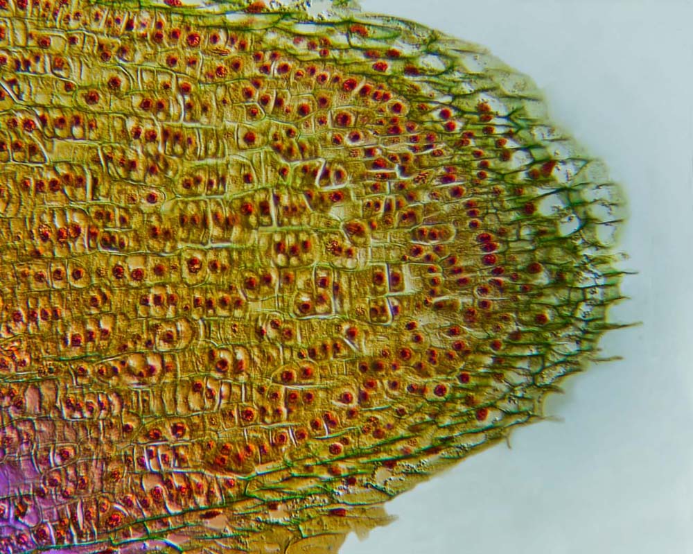
David Spears
David Spears Imaging, Taunton, Somerset, United Kingdom
Root tip section of a dicot plant (25.5x), Differential Interference Contrast
Mutant cell
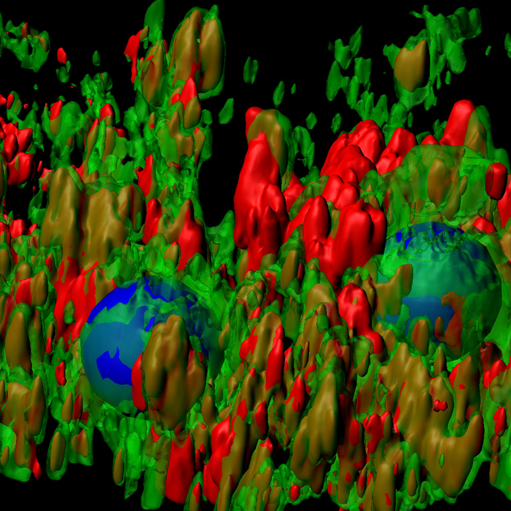
Dr. Donna Beer Stolz
University of Pittsburgh, Department of Cell Biology, Pittsburgh, Pennsylvania, USA
Mutant human alpha-1 antitrypsin aggregates (red) exiting the endoplasmic reticulum (green) in an iPS cell differentiated into a liver cell (200x), Confocal Reconstruction
Human muscle
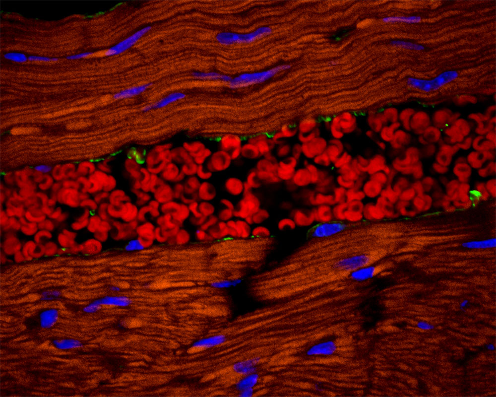
Dr. Tomasz Szul
University of Alabama at Birmingham, Department of Medicine/Pulmonary, Birmingham, Alabama, USA
Cross section of a human muscle tissue revealing red blood cells (erythrocytes) inside a blood vessel (600x), Fluorescence, Confocal
Mouse lung
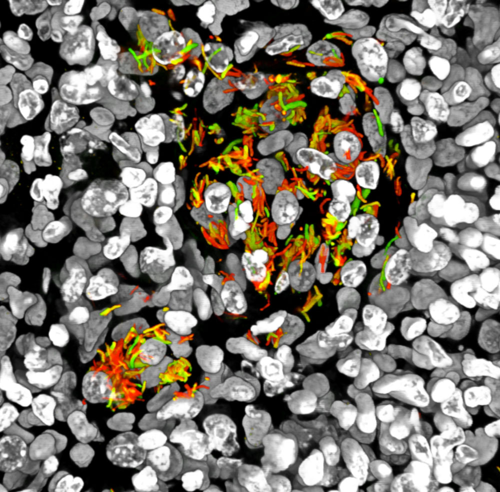
Dr. Shumin Tan
Cornell University, Department of Microbiology and Immunology, Ithaca, New York, USA
Lung tissue from a mouse infected with reporter Mycobacterium tuberculosis, engineered to fluoresce in response to environmental acidity and chloride concentrations (63x), Confocal
Twinned crystals
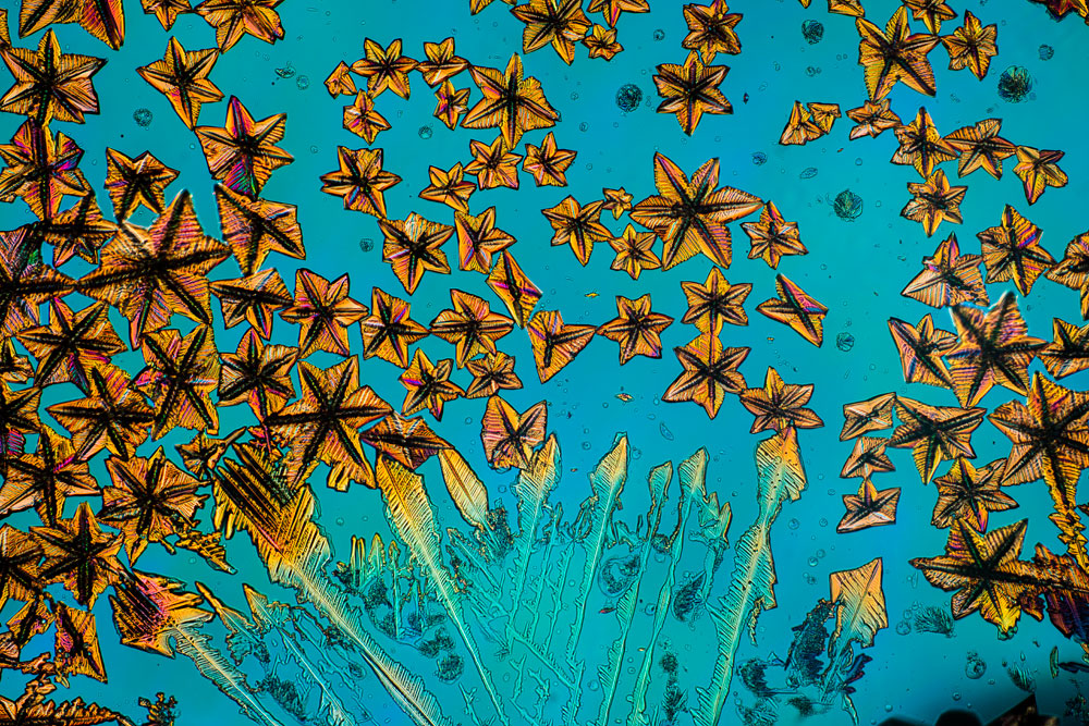
Dr. Ryoji Tanaka
Microphoto Studio "Cat's Glove," Ebina, Kanagawa, Japan
Twinned crystals of 4, 4'-dibromobiphenyl (25x), Polarized Light, Retardation Control
Living rotifer
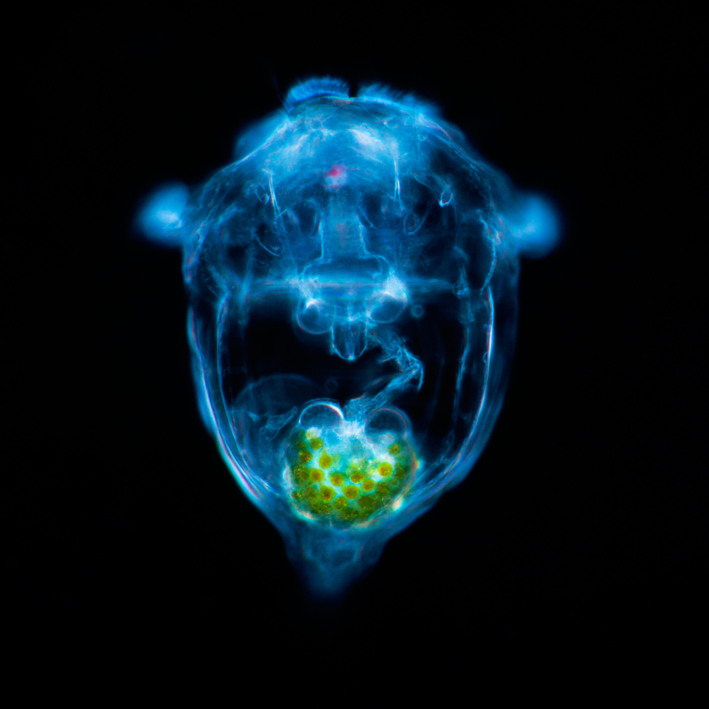
Dr. Bernd Walz
University of Potsdam, Potsdam, Brandenburg, Germany
Living rotifer (Synchaeta sp) (400x), Darkfield
Liquid crystal
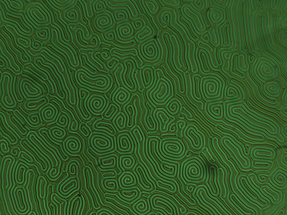
Dr. Giuliano Zanchetta
University of Milan, Department of Medical Biotechnology and Translational Medicine, Milan, Italy
Texture of a chiral thermotropic liquid crystal (20x), Polarized Light
Film
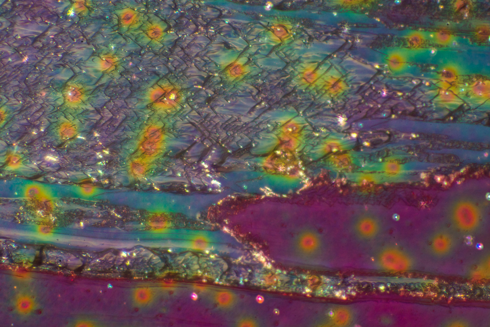
Teresa Zgoda
Rochester Institute of Technology, Rochester, New York, USA
Torn photographic film (Fujifilm) (10x), Reflected Light
Suspended particles
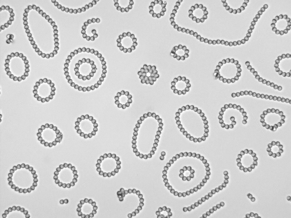
Jie Zhang
University of Illinois at Urbana-Champaign, Urbana, Illinois, USA
Janus particles (microparticles) suspended in water between two transparent electrode (40x), Brightfield

