Photos: Muscles and Bones Made with New 'Bioprinter'
A new 3D printer can print living tissue structures that could one day be used to replace injured or diseased tissue in patients.
"With further development, this technology could potentially be used to print living tissue and organ structures for surgical implantation," Dr. Anthony Atala, director of the Wake Forest Institute for Regenerative Medicine, who co-authored a study describing the new printer, said in a statement. [Read full story: 3D 'Bioprinter' Makes Replacement Bones, Ears]
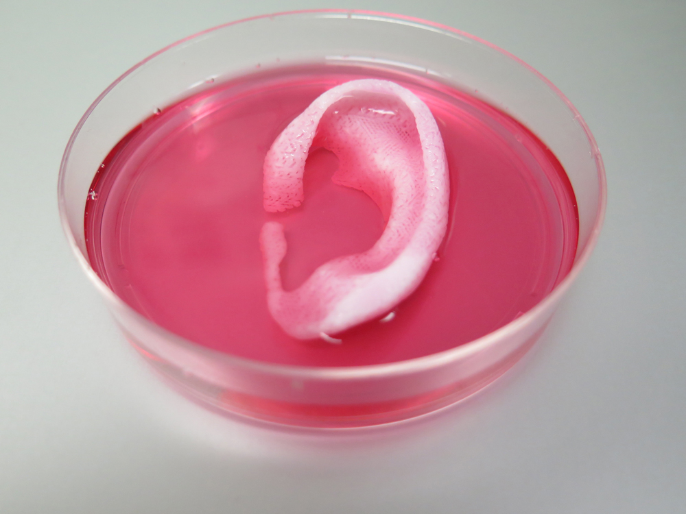
3D-printed ear structure
This photo shows an ear structure printed with the new bioprinter. In experiments, the researchers implanted such ear structures under the skin of mice to see if the structure tissue would survive. They found that the structures did survive, and had even developed blood vessels by two months after implantation, thanks to special microchannels printed throughout the structures. (Credit: Wake Forest Institute for Regenerative Medicine)
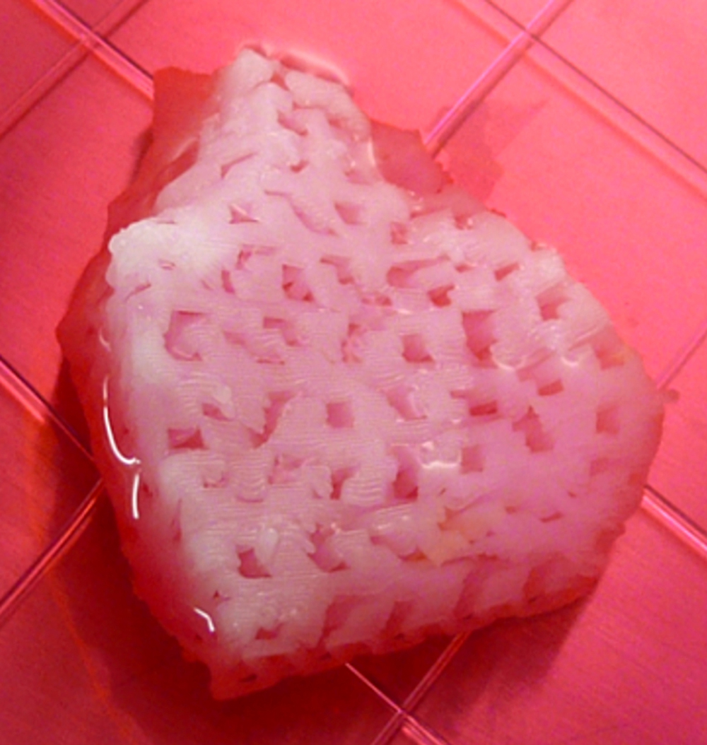
3D-printed jaw bone structure
This image shows a jaw bone fragment printed with the new bioprinter. The size and shape of the fragment corresponds to the size and shape of fragments that could be used for jaw reconstruction in human patients. (Credit: Wake Forest Institute for Regenerative Medicine)
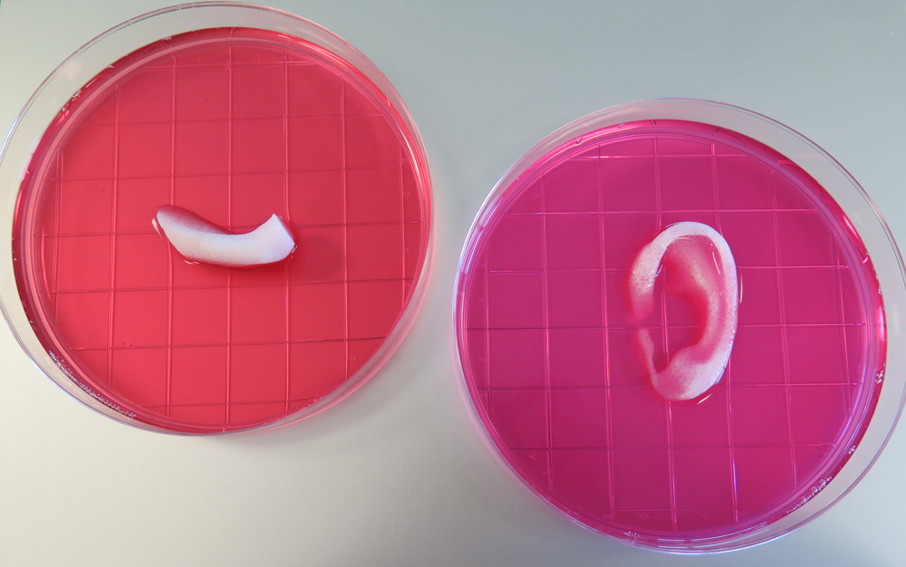
Tailor-made tissue
Get the world’s most fascinating discoveries delivered straight to your inbox.
This photo shows 3D-printed ear and jaw bone structures. The new printing system can use data from CT and MRI scans to tailor-make tissue for patients. For instance, if a patient is missing an ear, the printer could print a new matching ear structure based on a scan of their intact ear. (Credit: Wake Forest Institute for Regenerative Medicine)
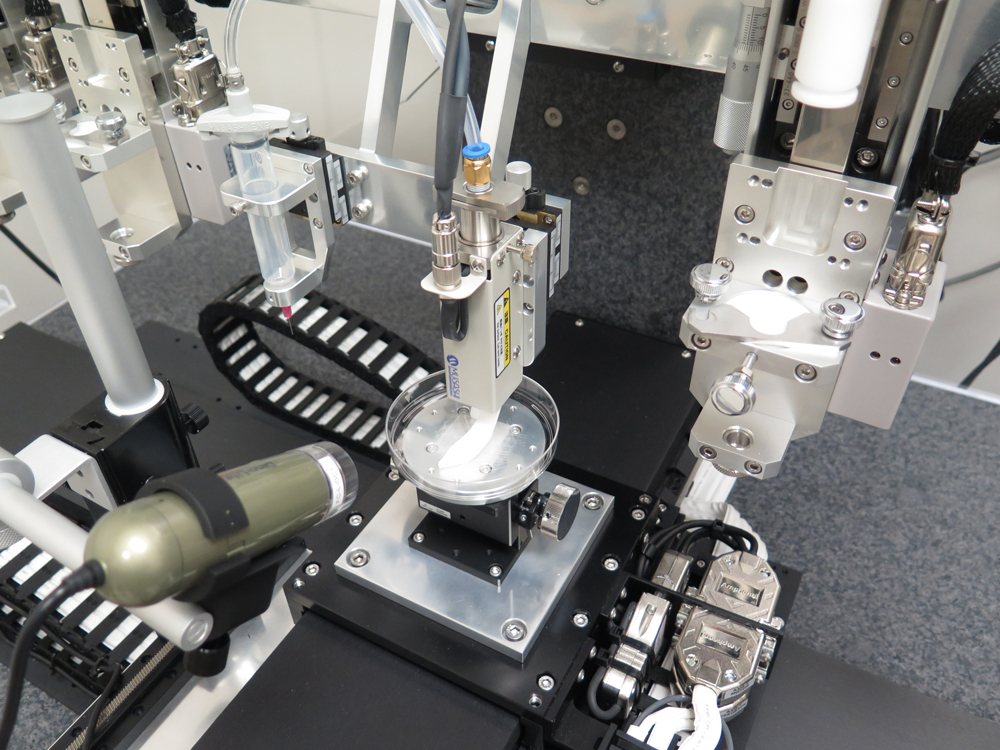
Printer at work
This photo shows the printing system at work, printing a jaw bone structure. The new printer deposits plastic-like materials to form the shape of the tissue and water-based gels that contain cells. This process allows the printed tissue to retain its shape and ensures the printing process does not damage the cells. (Credit: Wake Forest Institute for Regenerative Medicine)
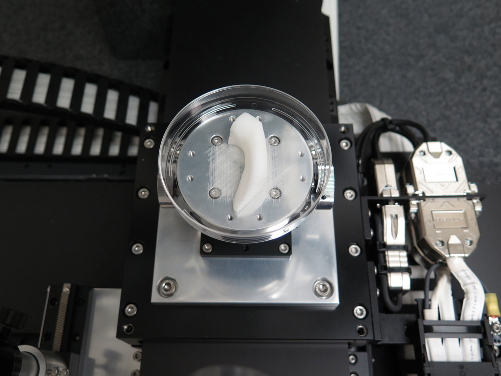
Larger, stronger tissues
This image shows a close-up view of the jaw bone structure during the printing process. The new printer allows researchers to print tissue and organ structures that are larger and stronger than the relatively simple and fragile tissues that researchers have engineered before. (Credit: Wake Forest Institute for Regenerative Medicine)
More research needed
This photo shows another bioprinted jaw bone structure. So far, the researchers have been able to implant only some of the tissue and bone structures they have made into rodents. Much more research is needed before these structures can be implanted in human patients, the researchers said. (Credit: Wake Forest Institute for Regenerative Medicine)
