Photos: The Secret Lives of Corals
Benthic Underwater Microscope
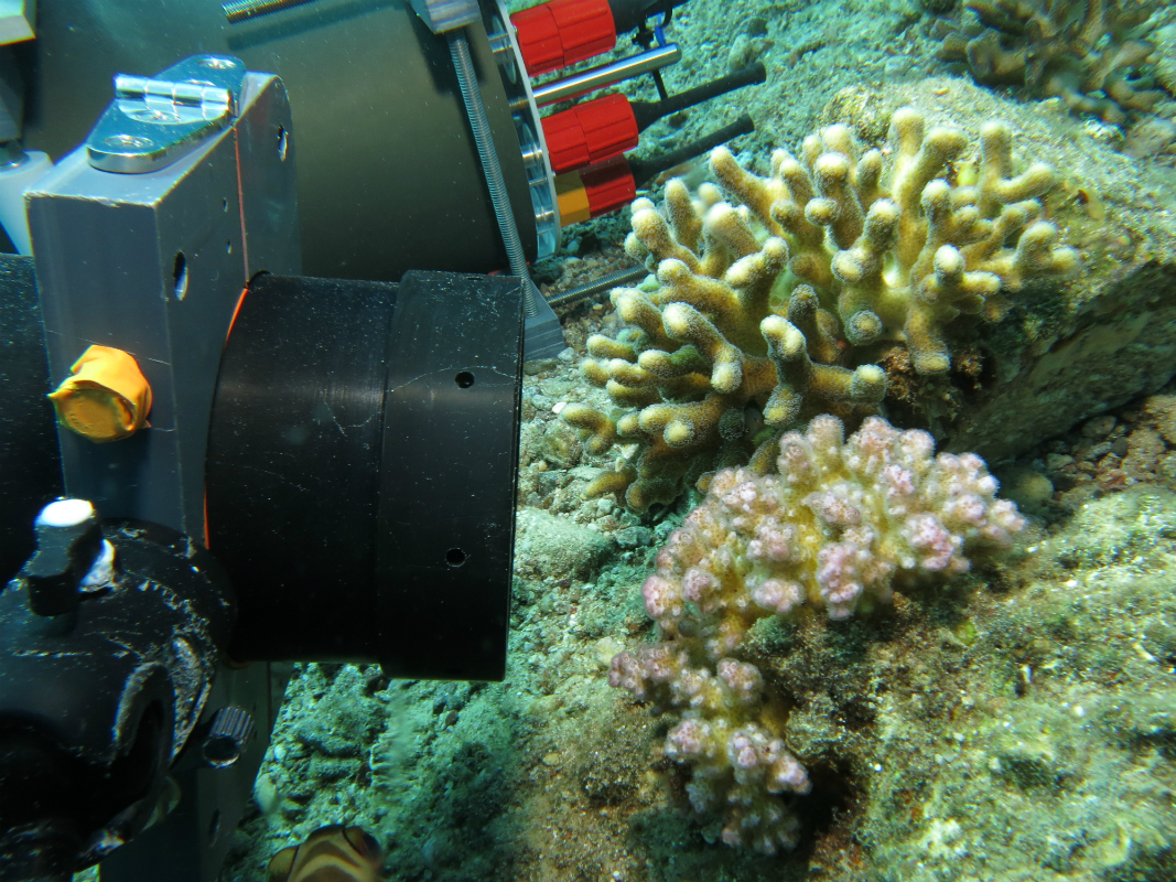
The Benthic Underwater Microscope (BUM) is positioned to study coral competition.
Stylophora
In situ image of the coral Stylophora taken using the 5x objective lens, in Eilat, Israel. The image is an enhanced DOF composite formed from a focal scan z-stack. The field of view is 1.7 x 1.4 mm.
Benthic Underwater Microscope image
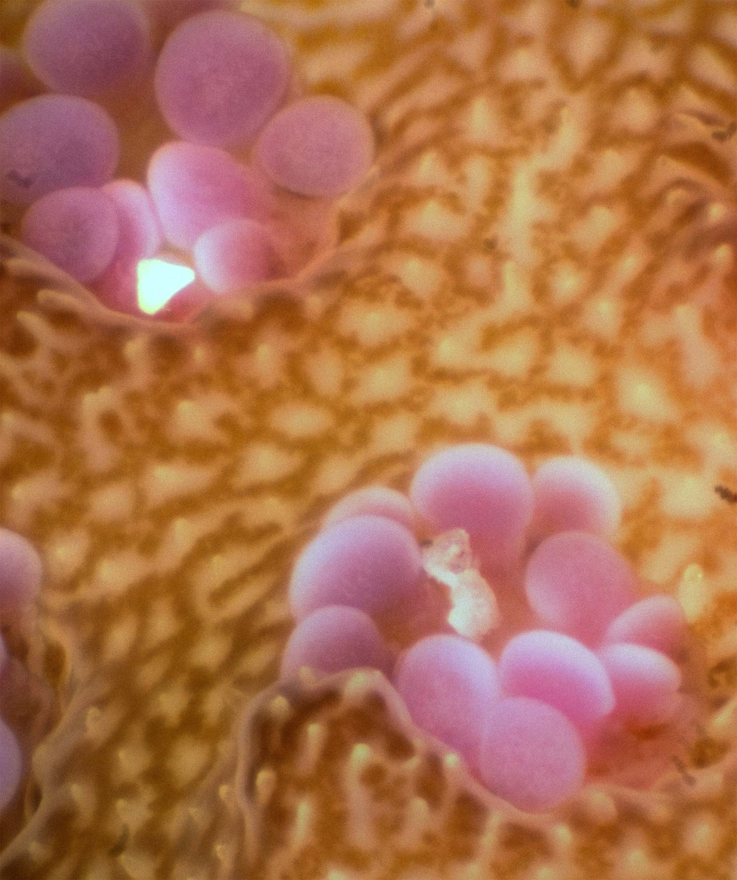
In situ image acquired using the 3x objective lens, in Eilat, Israel. The image is an enhanced DOF composite formed from a focal scan z-stack. The field of view is 1.7 x 1.4 mm.
Rhopalaea
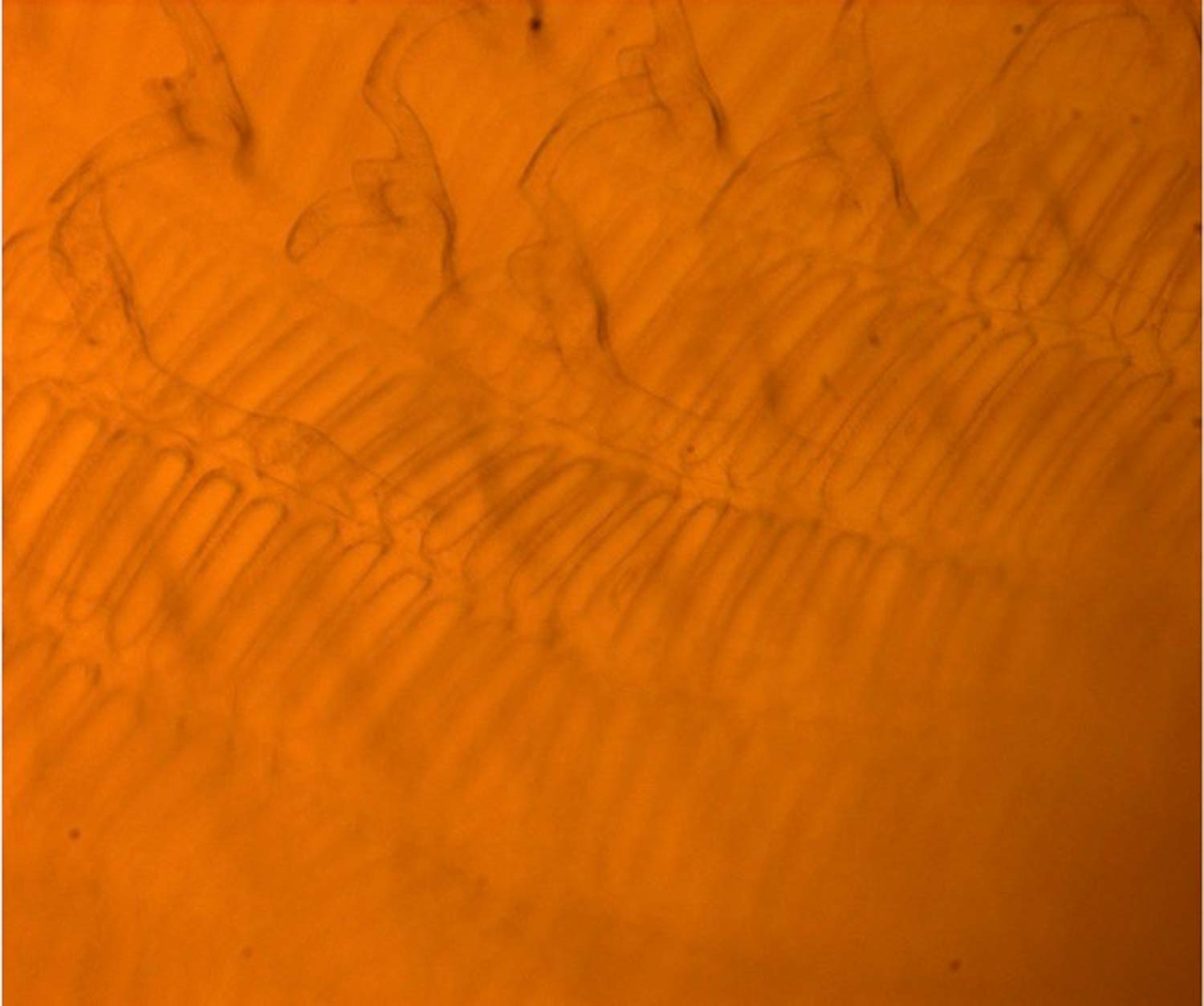
In situ image of the pharyngeal basket of the transparent ascidian Rhopalaea idoneta, taken using the 5x objective. Rhopalaea idoneta is a filter feeder which uses the mesh of the pharyngeal basket to capture plankton. The field of view is 1.7 by 1.4 mm.
First phases of bleaching
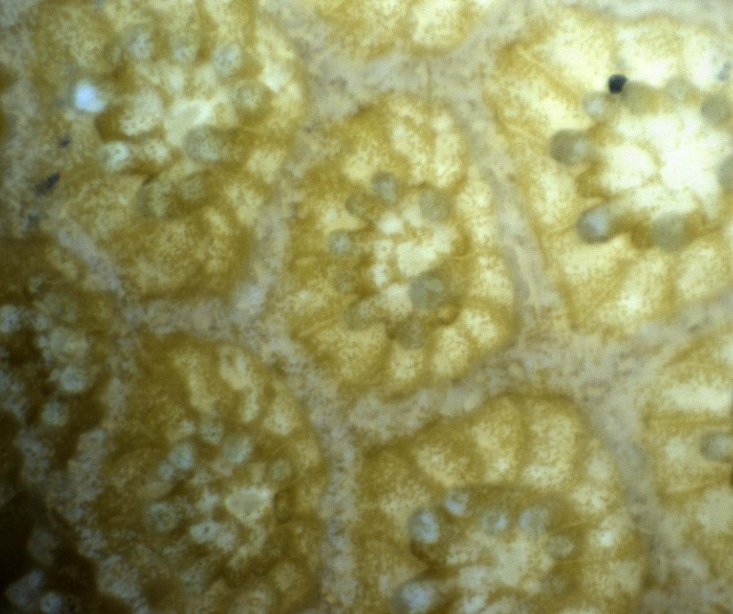
Porities coral with different levels of zooxanthellae loss. When few or no zooxanthellae can be seen in the coral the polyps have a translucent appearance.
More damage
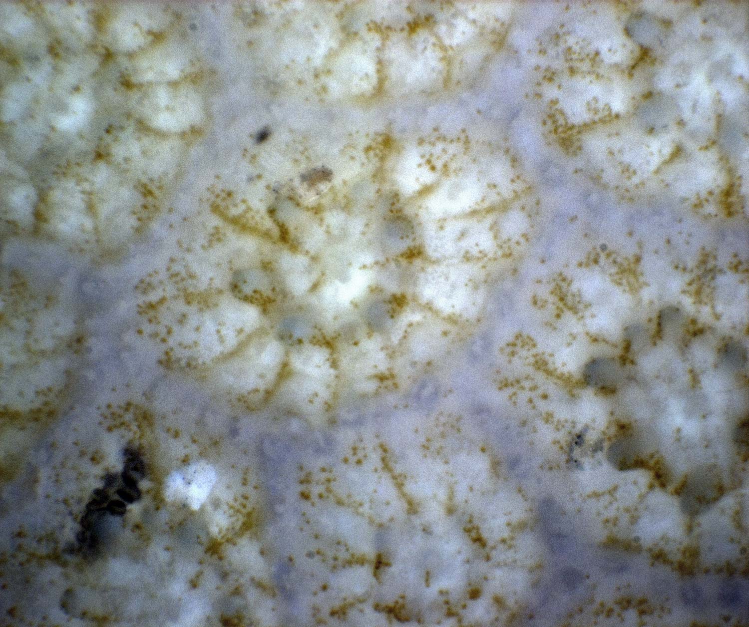
While translucent, the polyp structure and tentacles remain intact and visible, indicating that the polyp is still alive.
Beyond repair
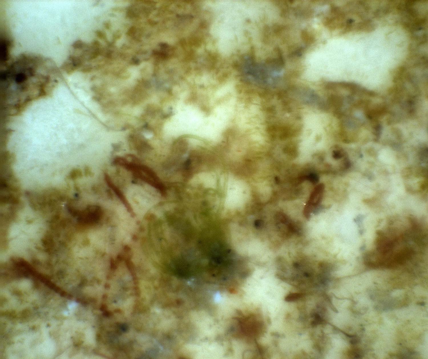
Bleached Porities polyps being colonized and overgrown by turf algae.
Sign up for the Live Science daily newsletter now
Get the world’s most fascinating discoveries delivered straight to your inbox.
Coral competition
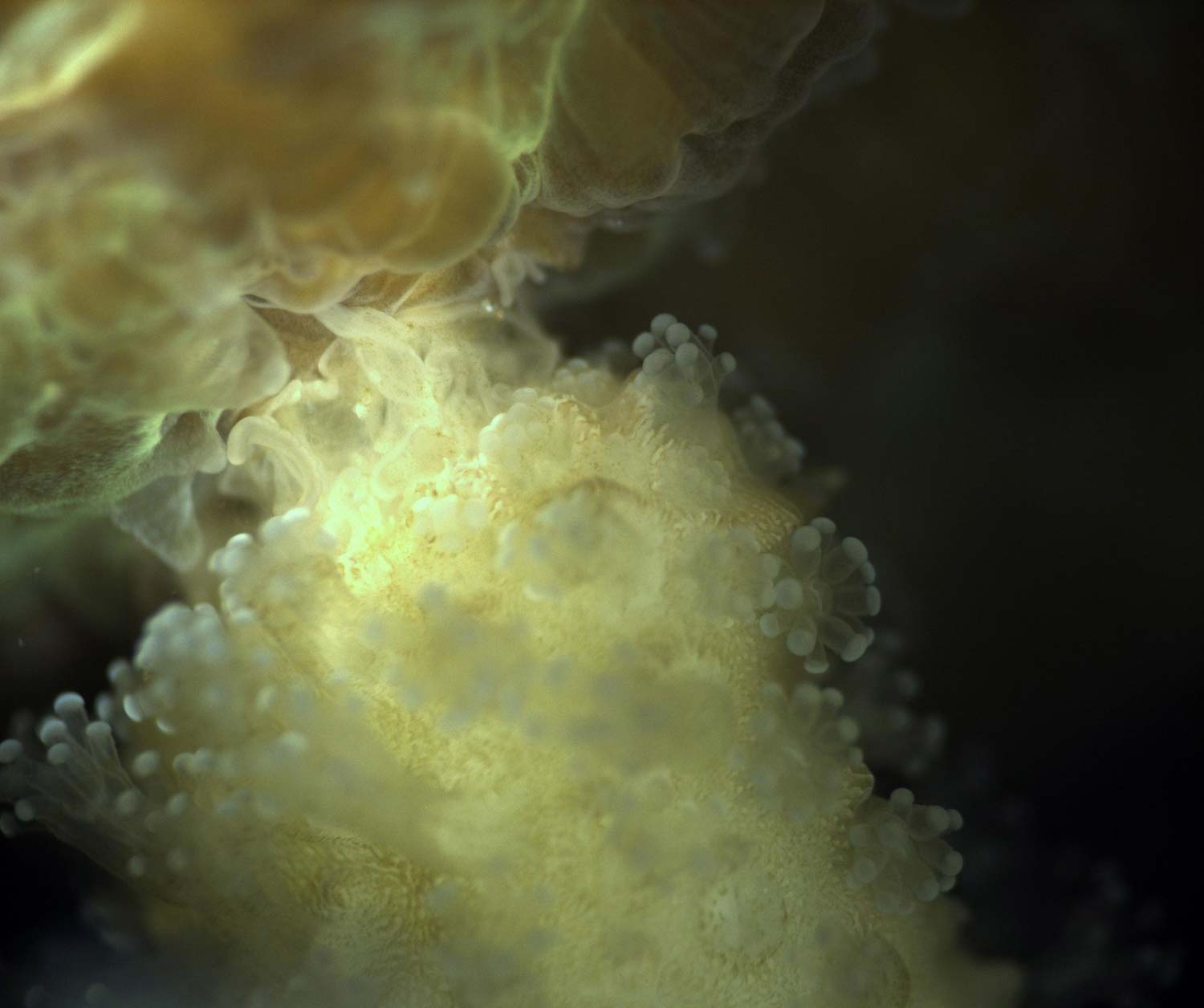
In situ image of competition between the corals Platygyra (left) and Stylophora (right) recorded in situ. The Platygyra emitting its mesenterial filaments. The field of view is 17 x 14 mm.
Pocillopora Coral Under White Illumination
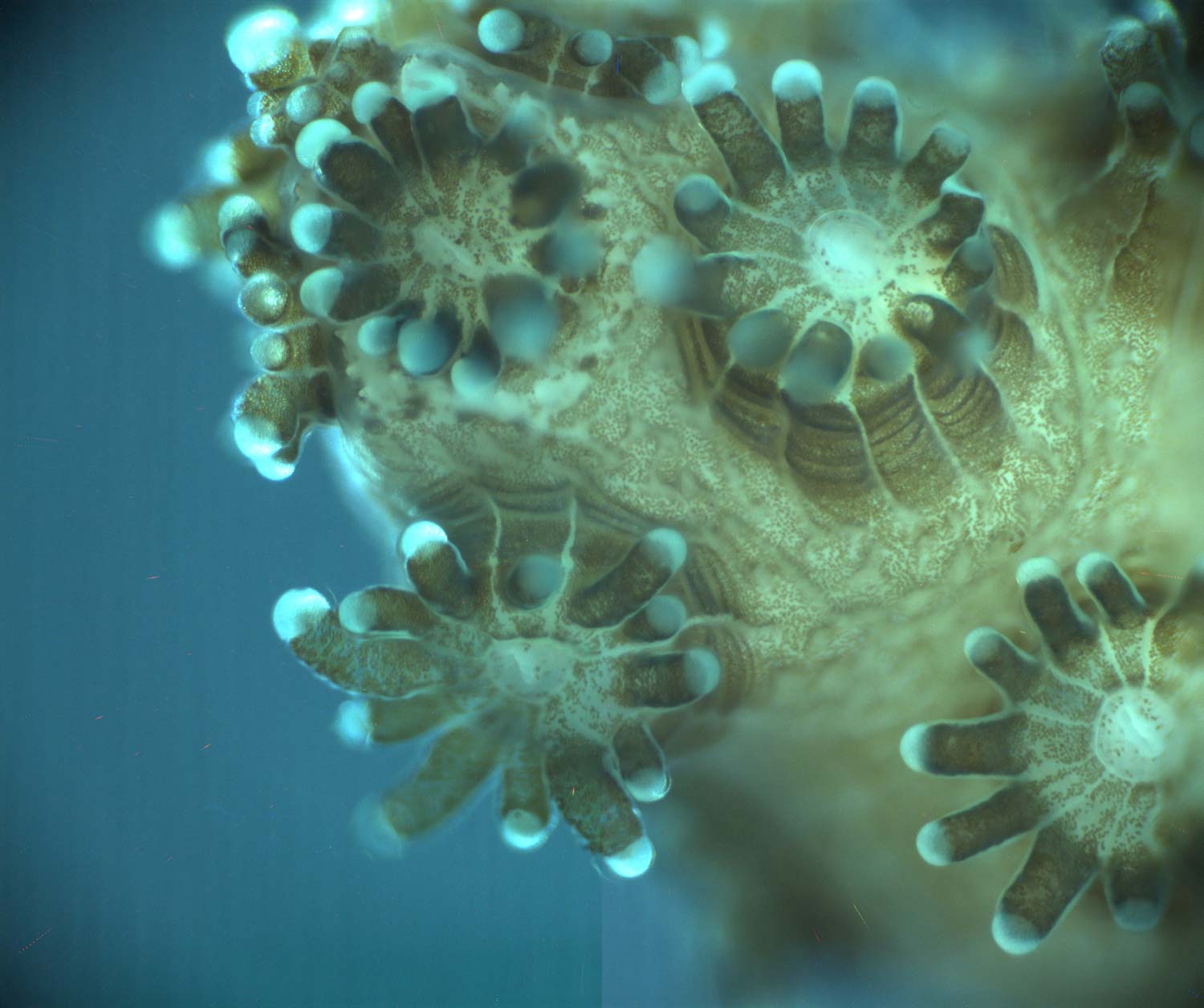
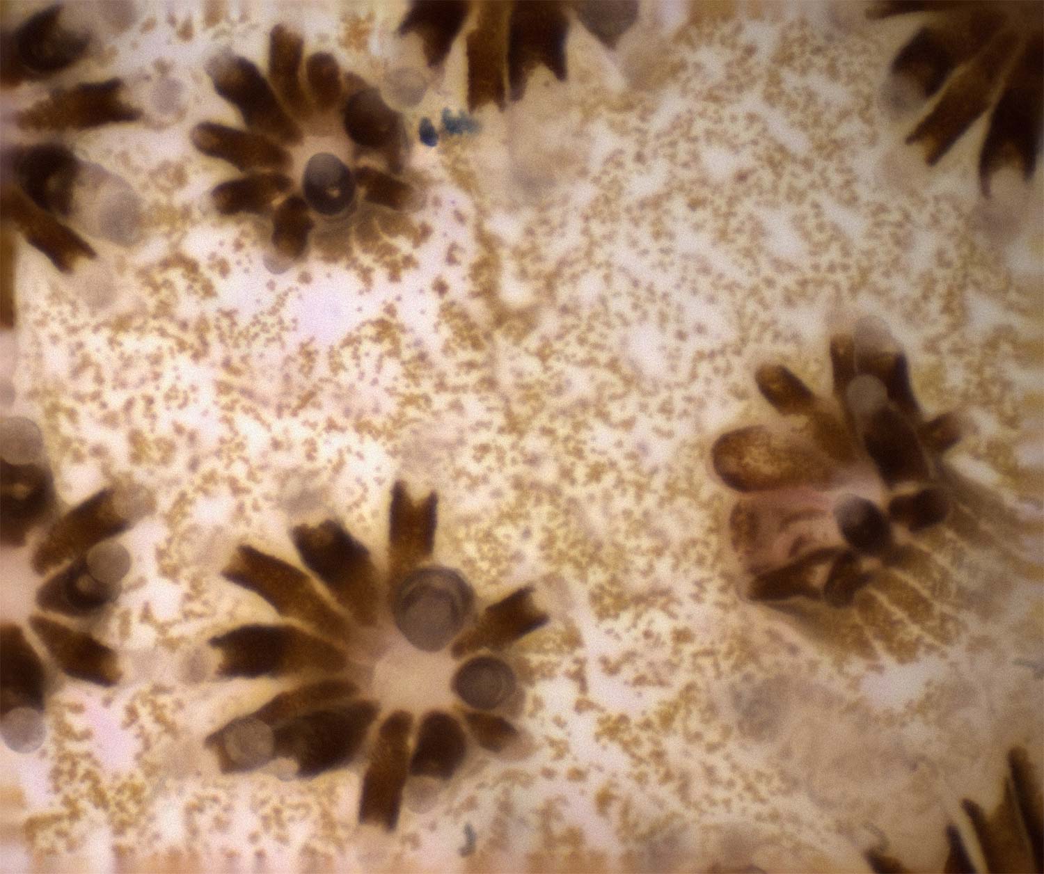
Image of the coral Pocillopora damicornis recorded in the lab. The image is an enhanced depth of field composite formed from several images taken at different focus planes. The small dots inside the polyps are individual zooxanthellae. The field of view is 4.2 x 3.5 mm.
Pocillopora Coral Competition
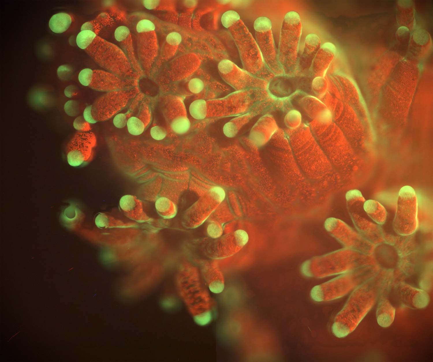
Fluorescent image of the coral Pocillopora damicornis taken in the lab. The image was formed by exciting the coral with blue light and using a filter to block any returning light in the blue portion of the spectrum. Two sources of natural fluorescence can be seen. Red fluorescence is produced by chlorophyll in single-celled symbiotic algae, Symbiodinium, living inside the coral. Green fluorescence is emitted from tips of the coral tentacles by a fluorescent protein naturally produced in the coral. The image is an enhanced depth of field composite formed from several images taken at different focus planes, the field of view is 4.2 x 3.5 mm.

