Peekaboo! Baby Brains Process Faces Just Like Adult Brains Do
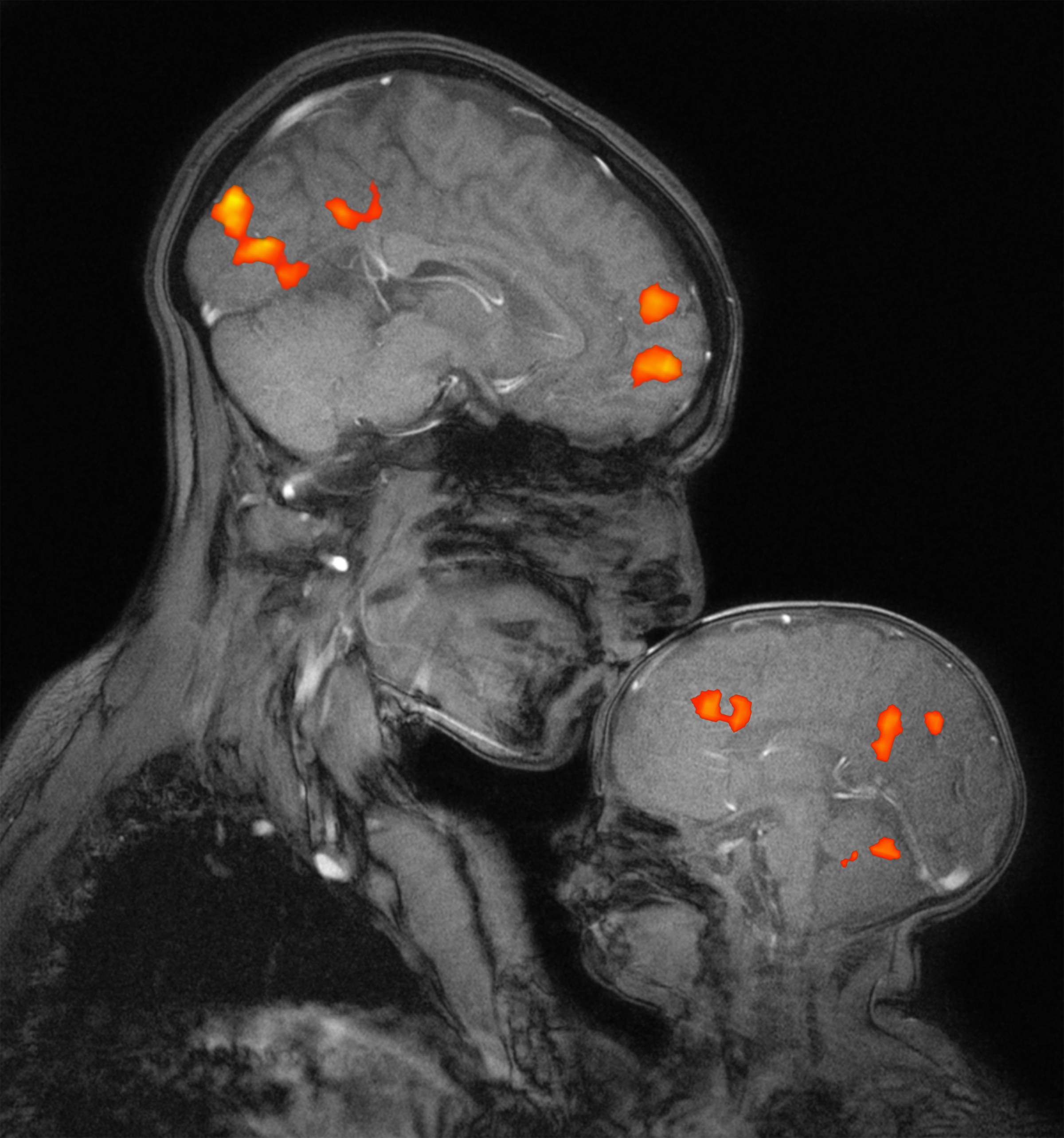
Babies as young as 4 months old process the faces and scenes that they see much like adults do, according to a new study. The findings suggest that the structure of the brain's visual cortex is already highly organized at birth or soon after.
The visual cortex is the part of the brain that processes all visual information. In adults, this area is highly compartmentalized into regions specialized to process certain kinds of objects, such as faces, houses or trees. Scientists have long wondered how the visual cortex got this way: Are these regions specified at birth, before the brain even knows what a face or tree looks like, or do they develop later as people grow and learn?
A definitive answer has eluded scientists, however, because of the inherent challenges of studying the brains of infants. The primary tool for studying these brain areas is the functional magnetic resonance imaging (fMRI) brain scan, which is challenging to use on infants. While the scan poses no radiation danger, subjects need to stay still and awake in the machine for several minutes at a time to produce a clear image. [10 Things You Didn't Know About the Brain]
Babies aren't exactly known for staying still, particularly when they are awake … and particularly when they are placed in the long tube of an MRI machine.
In what may be a feat more monumental than herding cats, researchers at the Massachusetts Institute of Technology (MIT) and Harvard Medical School have managed to capture crisp fMRI scans of nine squirming, gurgling, burping or otherwise unruly infants. The scientists employed various tactics to keep the kids relatively still, such as placing them in a specially designed infant seat and climbing into the MRI scanner with the babies to help them feel safe. [7 Baby Myths Debunked]
The scans revealed that the visual cortex of 4- to 6-month-old human infants is clearly spatially organized, with distinct regions responding preferentially to human faces versus natural scenes. Additionally, the results showed that the babies' responses to these images resembled those observed in adults. That is, brain activity extended throughout the cerebral cortex, from the front to back of the brain, the researchers wrote in their study, published today (Jan. 10) in the journal Nature Communications.
Neuroscientists hotly debate how much of the structure of the brain is specified at birth and how much of that structure arises from experience, said Rebecca Saxe, a professor of cognitive neuroscience at MIT who was the senior author on the study.
Sign up for the Live Science daily newsletter now
Get the world’s most fascinating discoveries delivered straight to your inbox.
"Some people think only the simplest, most basic functions are specified at birth … and everything else is learned from the pattern of experience," Saxe told Live Science. "Other people think lots of high-level 'cognitive' functions are supported by pre-existing machinery that is 'ready to go' at birth in one way or another." [11 Facts Every Parent Should Know About Their Baby's Brain]
The answer may lie somewhere in between, according to Saxe's project with infants, which was led by Ben Deen, then a graduate student in Saxe's lab and now a postdoctoral fellow at the Rockefeller University in New York. The team found that the visual cortices of infants and adults are not identical, though they are similar, suggesting that the structure becomes refined through development, the researchers said.
Still, the new findings support the hypothesis that specification in the brain at birth provides a sort of scaffolding that ultimately leads to precise category-selective brain regions in adults, the researchers said. As another example, Saxe cited the part of the brain called the "visual word form area" where humans process alphabets and ideograms. Reading is a relatively new phenomenon in the human experience; for all humans to possess such a specified brain area for visualizing words, it must be built upon innate scaffolding.
In a TEDx talk in June 2016, Saxe explained that she turned to studying infant brains to better understand the origin of the human mind. She wanted to answer questions like, "What is innate, what is learned, and how much of what people think about the world is universal?" Her own newborn boy was one of her first test subjects in her group's fMRI project.
Follow Christopher Wanjek @wanjekfor daily tweets on health and science with a humorous edge. Wanjek is the author of "Food at Work" and "Bad Medicine." His column, Bad Medicine, appears regularly on Live Science.

Christopher Wanjek is a Live Science contributor and a health and science writer. He is the author of three science books: Spacefarers (2020), Food at Work (2005) and Bad Medicine (2003). His "Food at Work" book and project, concerning workers' health, safety and productivity, was commissioned by the U.N.'s International Labor Organization. For Live Science, Christopher covers public health, nutrition and biology, and he has written extensively for The Washington Post and Sky & Telescope among others, as well as for the NASA Goddard Space Flight Center, where he was a senior writer. Christopher holds a Master of Health degree from Harvard School of Public Health and a degree in journalism from Temple University.
