Lifelike Models with 'Working Organs' Help Doctors Hone Surgical Skills
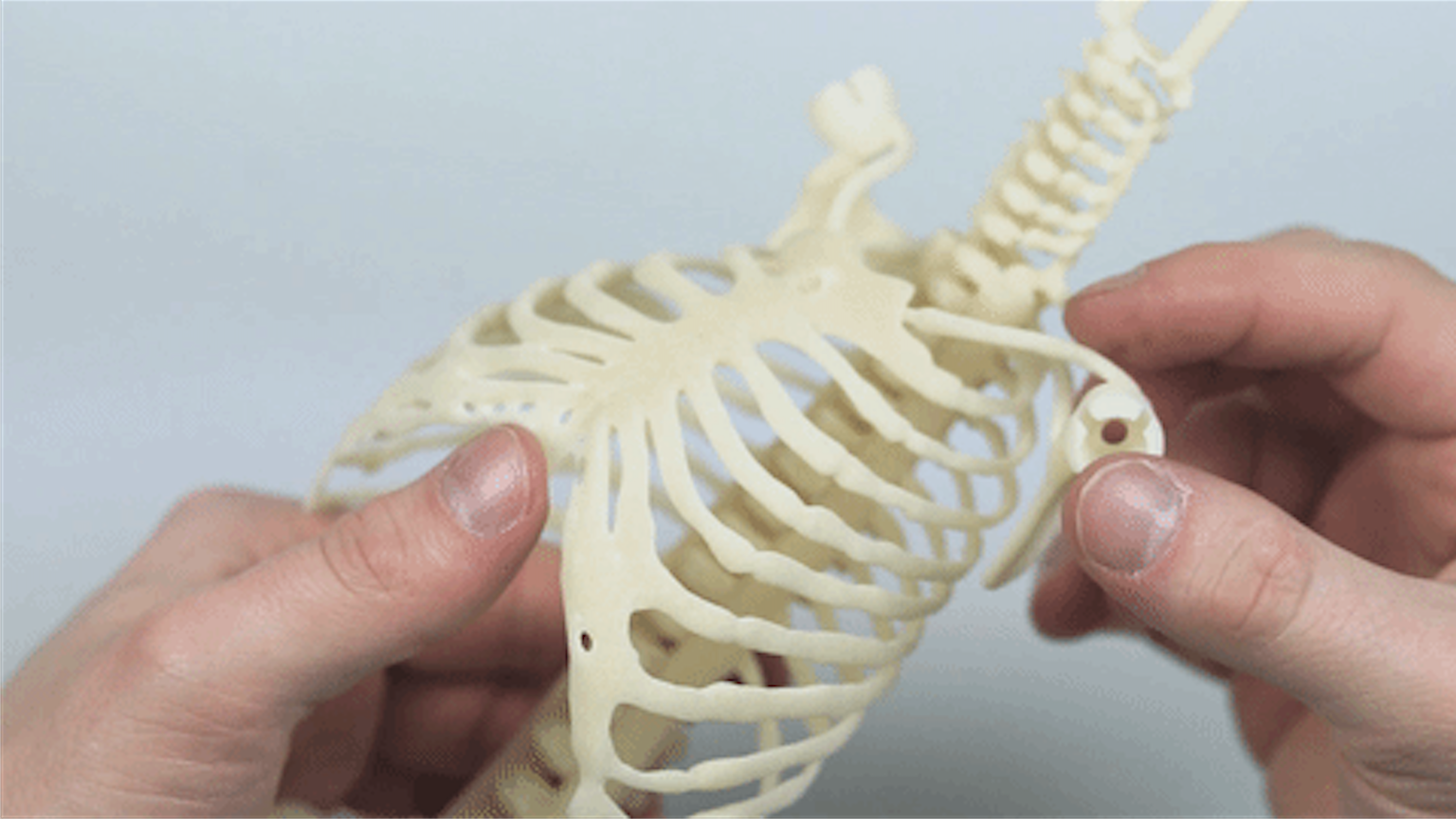
New, lifelike models of newborns that are as squishy as real babies and even include mimics of working organs could help surgeons and nurses train to perform life-saving procedures, researchers say.
The baby mimics were created using 3D printing and were designed to better replicate the anatomical complexity and feel of real newborns. This is important because doctors usually hone their surgical skills on life-size manikins.
And the more realistic the model, the better.
Currently, the manikins that are available to doctors "tend to feel very mechanical, rather than biological," said Mark Thielen, a medical design engineer at the Eindhoven University of Technology in the Netherlands. [5 Amazing Technologies That Are Revolutionizing Biotech]
For example, instead of a replica of a rib cage inside a manikin, there's usually "a central spring with a plastic plate on top, covered by rubber skin," Thielen said.
To make manikins of newborns more realistic, Thielen and his colleagues turned to 3D printing. Their 3D-printed manikins open up "the possibility of teaching and investigating clinical procedures to an extremely high level of accuracy and understanding," Thielen told Live Science.
3D printers make items by depositing layers of material just as ordinary printers lay down ink, except 3D printers can also layer materials on top of each other to build 3D objects. These creations are typically made of varieties of plastic or resin.
Sign up for the Live Science daily newsletter now
Get the world’s most fascinating discoveries delivered straight to your inbox.
"Without 3D printing, this work would have been impossible," Thielen said in a statement. "The sheer complexity of human anatomy is very hard to re-create realistically with any other production method, not to mention the cost and time differences."
Making the models
To build the most lifelike model possible, Thielen and his team worked from the inside out, starting with the organs.
Data from MRI scans of newborns was used to develop the 3D models for the manikins' organs. The realistic organs were printed with the help of 3D Hubs, an online network that connects people to 3D printing services. The goal was not only for the organs to look and feel like their real counterparts, but for them to function like real organs, too. For example, the 3D-printed heart had highly detailed, working valves. In the future, Thielen and his team hope to design model organs that function as similarly to real organs as possible.
Furthermore, there are no springs and plastic plates in these manikins. Rather, the printed organs are housed in manikins with 3D-printed rib cages and spines. To mimic blood, fluid is injected into tubes in the manikins. [7 Cool Uses of 3D Printing in Medicine]
"The aim is to provide a high level of realistic tactile feedback when performing clinical interventions on them," Thielen said. In other words, when the surgeons move a part of the manikin or apply pressure to a certain area, it feels and moves like the real thing.
In addition, the manikins have a variety of sensors embedded within them to measure movements such as bending and sliding, as well as to gauge pressure in different areas and the flow of fluids. These sensors can make measurements such as the angle at which the head is tilted backward during a procedure, the amount of air squeezed into the lungs or the amount of blood pushed out of the left and right sides of the heart during chest compressions, Thielen said.
Thielen plans to feed data from the sensors into computer models of the human body to predict how operations performed on the manikins might affect a real patient when performed in real life.
Although the manikins are still in development, the initial tests looked promising, Thielen said. If the newborn-baby manikins are successful, future work could focus on the development of more realistic manikins of adults and adult-sized organs, he said.
"I believe that developing and advancing what we started here can aid medical research in a broader scope," Thielen said. "We could potentially create realistic patient models of other body parts to strengthen medical training for emergency procedures and pregnancies."
Original article on Live Science.

'A relationship that could horrify Darwin': Mindy Weisberger on the skin-crawling reality of insect zombification
'Dispiriting and exasperating': The world's super rich are buying up T. rex fossils and it's hampering research
Trove of dinosaur footprints reveal Jurassic secrets on Isle of Skye where would-be Scottish king Bonnie Prince Charlie escaped

