Fetus Brought Partway Out of Womb for Tumor Surgery

Surgeons in Philadelphia recently removed a tumor from what may be the tiniest heart ever to undergo surgery. The heart belonged to a 21-week-old fetus, still inside the womb.
"The fetus was just about 6 inches [15 centimeters] in total length, and his heart was the size of a peanut, perhaps a centimeter or less," said Dr. Jack Rychik, director of the Fetal Heart Program at Children's Hospital of Philadelphia (CHOP).
Doctors at CHOP have performed more than 1,400 fetal operations since 1995, according to the hospital's officials. Most of those procedures have been done to remedy cases of spina bifida, a birth defect of the spine.
Operating on a fetus's heart, however, is extremely risky and rarely done, Rychik said. He told Live Science the CHOP team undertook the procedure only because it was a matter of life or death for the fetus. The tumor was the size of a walnut, larger than the heart itself, and was squeezing the tiny organ, Rychik said.
The mother, Cecilia Cella, lives in Uruguay. She had gone to see a pediatric cardiologist there, Dr. Roberto Canessa, who texted Rychik with video of a strange mass he saw on Cella's sonogram. [7 Baby Myths Debunked]
"He sent it to me and said, 'What the hell is this?'" Rychik said.
Rychik said he recognized the mass as an extremely rare, rapidly growing tumor called an intrapericardial teratoma. Such tumors grow on the sac surrounding the heart and can place deadly pressure on a fetus's heart if left unchecked.
Get the world’s most fascinating discoveries delivered straight to your inbox.
Fortunately, Cella and her partner, Pablo Paladino,were able to fly to Philadelphia in a matter of days, and the CHOP team readied for surgery.
"Had we waited another day, I think it would have been too late," Rychik said. "The tumor was just too large."
To operate on the fetus, the doctors first placed the mother under general anesthesia, which anesthetizes both mother and the fetus. The operating team included lead surgeon Dr. Holly Hedrick, Rychik and three other surgeons.
Then, the surgeons made an incision into the mother's uterus and drained the amniotic fluid (it was later replaced with artificial amniotic fluid). At this point, the fetus, a male, was further anesthetized, to ensure it would remain still for the procedure.
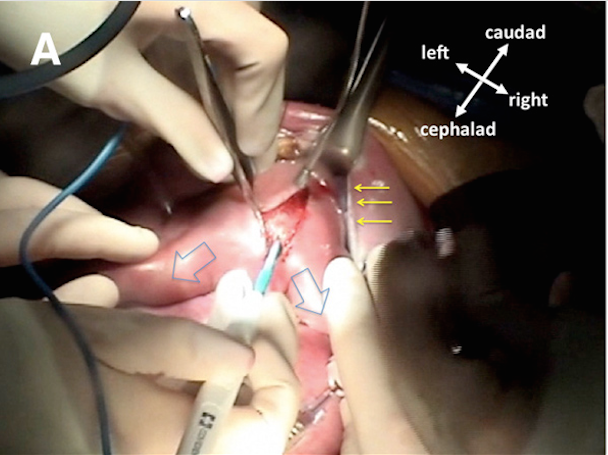
Then, it was a matter of gaining access to the fetus's heart. To do this, Rychik explained, the team delicately lifted the fetus's arms out of the uterus, leaving the head and rest of the torso inside the womb.
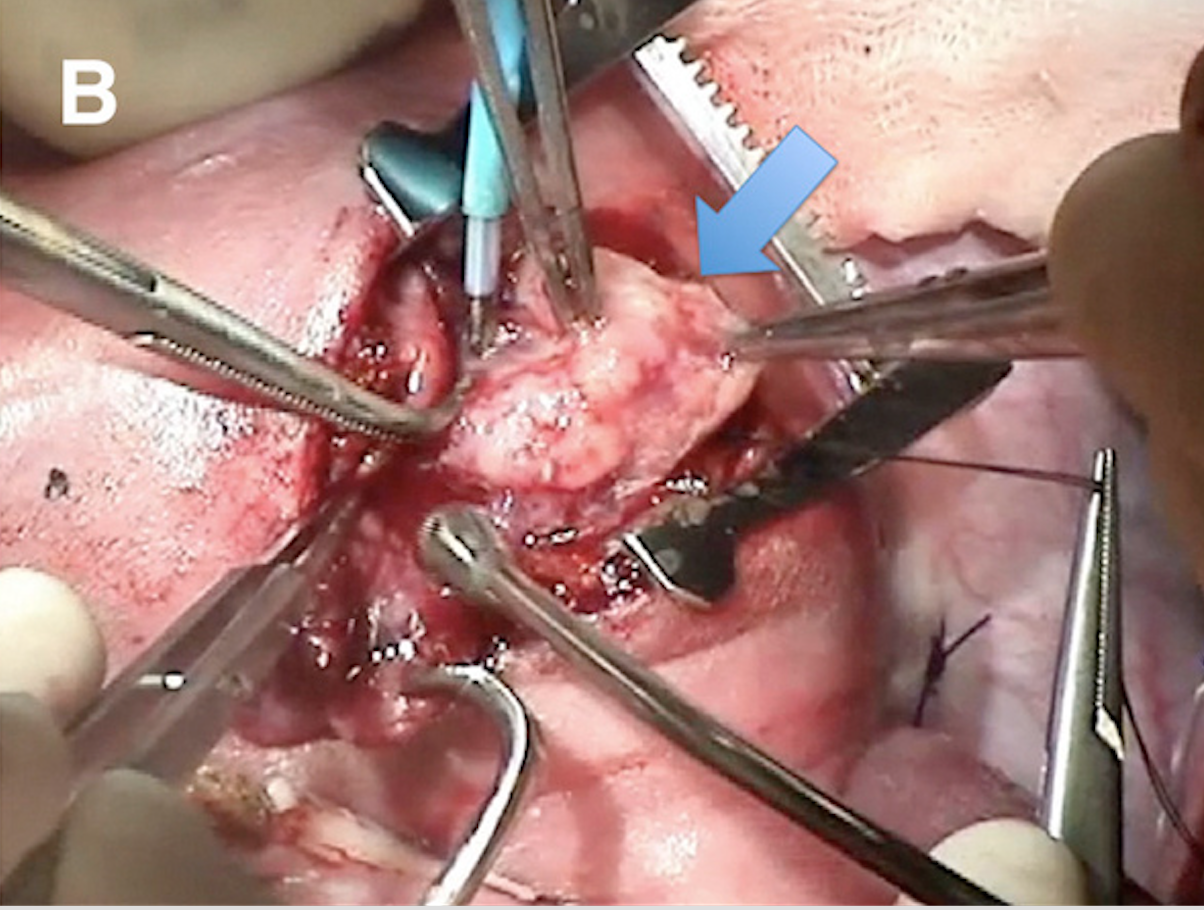
"By bringing the arms out and the chest up, [we ensure that] the chest is available to the surgery," Rychik said. "Then, an incision is made into the chest and the ribs are cut as they would be in an adult."
The doctors were able to excise the tumor from the fetal heart's sac. They then sewed up the incisions in the fetus, put it into the womb and sewed up the uterus, so the pregnancy could continue. Cella was then sewn back up.
While the procedure was underway, the team constantly monitored the fetus's heart through images projected by a very tiny ultrasound probe, which Rychick guided. The live sonogram images helped the surgeons avoid compressing the heart during the procedure and blocking blood flow.
The 3-hour procedure was mostly a success, but the surgeons weren't able to cut away all of the tumor, Rychik said. About 2 percent of the tumor was "too intimately attached" to the heart, he said.
"We worried if we removed that last bit, it may have caused damage to the heart," he said.
Three weeks after the surgery, the tumor had started to grow and appeared again on sonograms. Nonetheless, Rychik said the surgery bought the fetus vital additional weeks inside the mother's womb.
The baby, named Juan, was born on Dec. 11, 2016, at 31 weeks. Two weeks later, Juan underwent another surgery to remove all remaining traces of the tumor. He is now nearly 4 months old, back home in Uruguay and reportedly doing well.
Before Juan, the CHOP team had previously performed an in utero heart surgery like this just once, on a 24-week-old fetus, who is now a healthy 3-year-old thriving at his home in Vermont. [Social Surgery: Images of Live-Tweeted Operations]
The doctors wrote about that case, along with seven other fetal heart-tumor cases that they saw from 2009 to 2015 in a report they published in December 2016 in the American Journal of Obstetrics and Gynecology. In six of those cases, the doctors could not operate and the fetuses died. In one case, the doctors were able to deliver the baby early and perform the surgery after delivery.
The one case that involved a successful in utero surgery was that of the now-3-year-old Tucker Roussin, who loves sandboxes and monster trucks, the Philadelphia Inquirer reported at the time. Juan and Tucker are now the only two known fetuses to have undergone the procedure.
"Until now, we've only been able to watch these tumors grow and inform the mother that the fetus probably would not be able to survive," Rychik said. "Now, we're showing that a different result is possible."
Originally published on Live Science.


