Water Flea Giving Birth Makes a Big Splash in 'Small World' Videos

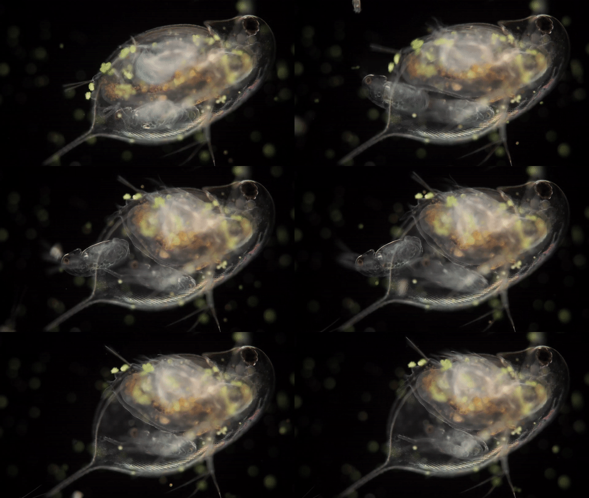
Giving birth has never looked as easy (and weird) as it does in a video captured by photographer Wim van Egmond. In it, a wee see-through daphnia, or water flea, expels a wriggling, googly-eyed larva, its body just as transparent as its mama's. Seconds after emerging into the water surrounding its mother, the young water flea darts swiftly away.
Van Egmond's footage earned a top spot, along with other mesmerizing videos, in the annual 2018 Nikon Small World in Motion contest, now in its eighth year.
Today (Sept. 27), Nikon representatives announced this year's five winning videos and 18 that received honorable mentions for exceptional footage of activity in the natural world that's too small to be seen with the naked eye. During 2018, contenders for Nikon recognition trained their microscope lenses on fragile and tiny subjects such as coral polyps, aquatic worms, bacteria and hatching insect eggs, all of which are nothing short of remarkable when seen up close.
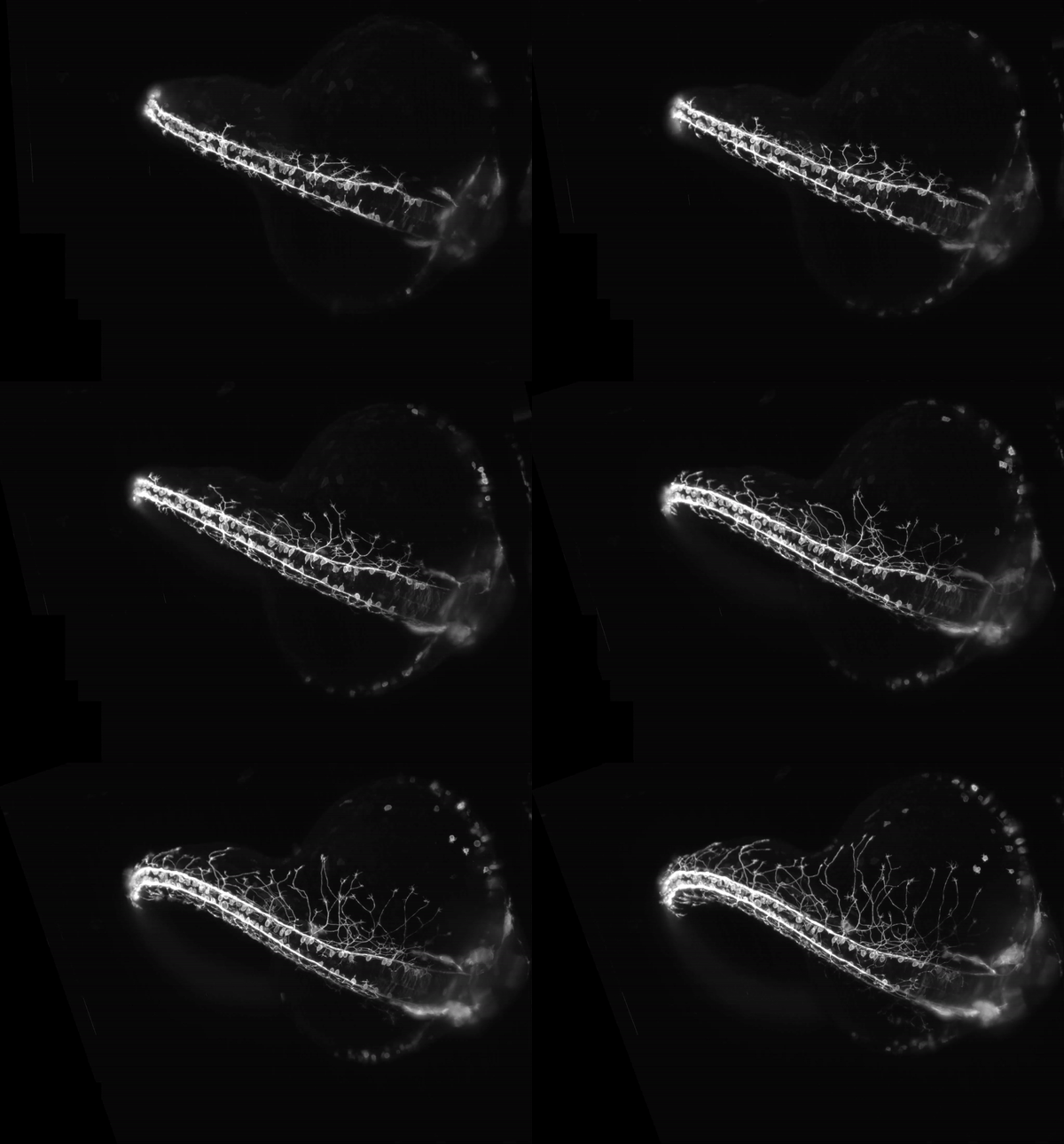
But only one video could be awarded first place, and that honor went to an astonishing time-lapse of a zebrafish embryo's growing nervous system. Elizabeth M. Haynes and Jiaye "Henry" He, researchers with the Department of Integrative Biology at the University of Wisconsin-Madison, recorded 16 hours of embryonic development, capturing the delicately branching filaments as they extended gracefully from the spinal column. [Magnificent Microphotography: 50 Tiny Wonders]
Scientists studying zebrafish embryos typically suspend them in gel blocks, but gel would have stunted the neurons' growth, Haynes told Live Science in an email. So, Haynes and He instead placed the embryo in water, capturing the unimpeded growth cycle of the developing neurons.
However, this posed a different challenge, as the embryo could easily have drifted away from the microscope lens during the long hours while they were filming it, Haynes said.
"There was an amount of luck present for it to remain in a good position during the entire movie," she explained.
Get the world’s most fascinating discoveries delivered straight to your inbox.
Lengthy exposure to light can also damage delicate embryos. But the researchers solved this problem with a special technique called Light Sheet Fluorescence Microscopy, which exposed the embryo to a much lower amount of laser power, "keeping it happy and healthy," Haynes said.
"This type of scope can also collect images extremely fast, which is another benefit. Without this technology, there’s no way we could have acquired such a remarkable movie," she added.
Imaging and examining growing neurons in zebrafish embryos could help researchers who study neuron growth and function in people, and may lend insights to their investigations of neurodegenerative disorders such as Alzheimer's disease, according to a statement issued by Nikon.
"Being able to see development happening in concert like this allows us to understand the big picture much better, and see things we hadn’t even considered looking at before," Hayes said.
'Clear, crisp and captivating'
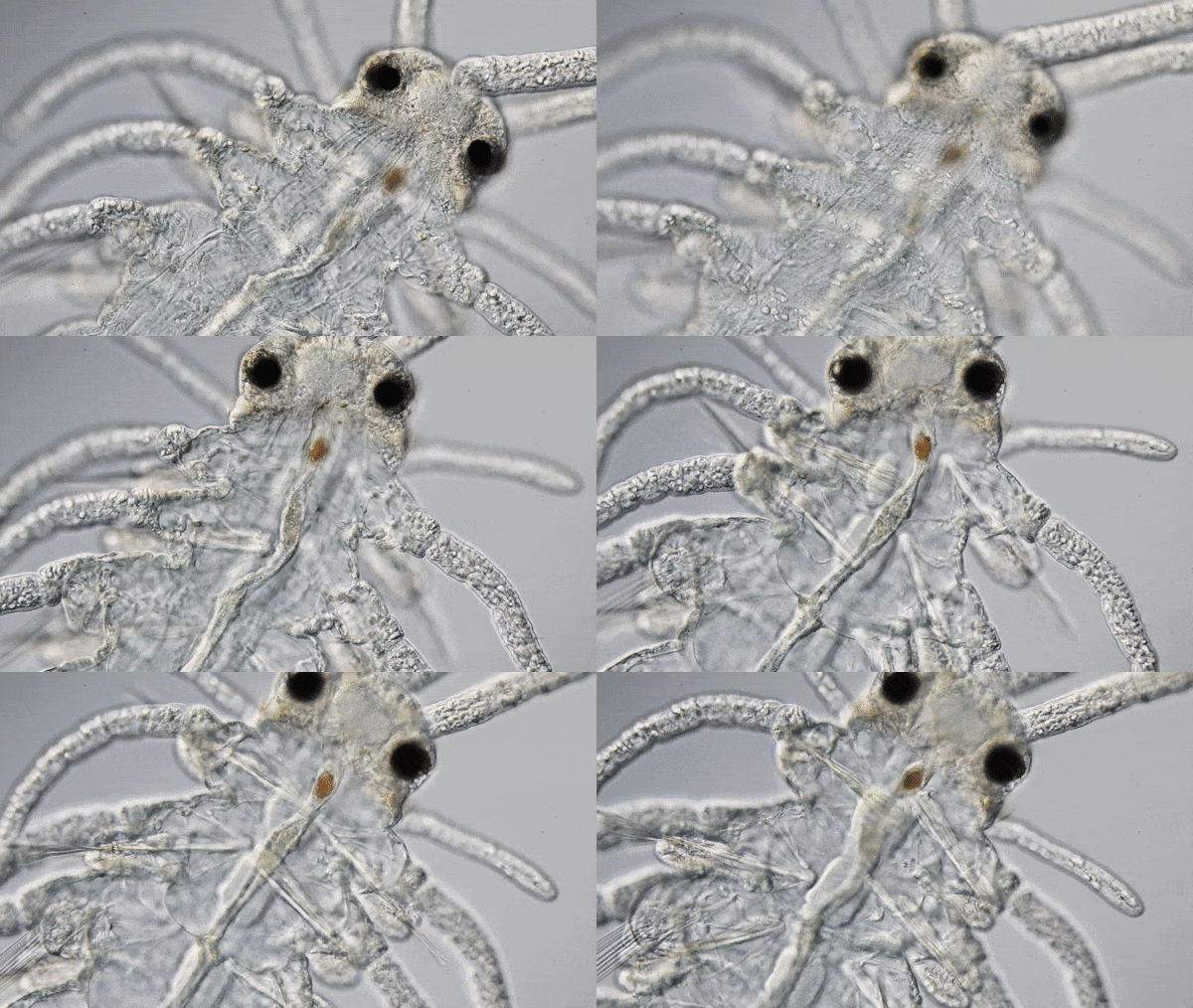
A video that earned third place shows a transparent bristle worm, revealing rigid internal structures that churn rapidly as the worm digests a meal. Filmed by Rafael Martín-Ledo, a marine-invertebrates researcher with the Ministry of Education, Culture and Sports in Spain, the video shows that the worm's movements displace a large blood vessel in its back.
Van Egmond, of the Micropolitan Museum in the Netherlands, has been recognized by Nikon Small World more than 30 times, according to the contest website. But what makes a video stand out from the crowd? Judges look for a number of qualities, including technical excellence, topical interest, and imagery that is "clear, crisp and captivating," contest judge Tristan Ursell, an assistant professor of physics with the Institute of Molecular Biology at the University of Oregon, told Live Science in an email.
"Many of the most common mistakes are the same mistakes that happen in regular photography and videography," Ursell explained. "Images may be out of focus, they may drift through time or not be long enough to convey a compelling visual story, [or] their contrasting might make important features either too dim or washed out," he said.
"And because microscopy deals with the very small, often even if everything else is perfect, it can be difficult to resolve small and/or fast moving objects and organisms," Ursell said.
Previous winners of the Nikon competition include video of a growing plant root, beads of sweat oozing between grooves in a fingerprint, and a starfish larva surrounded by swirling water vortices that it stirred up with cilia to find food.
These incredible visuals serve as a reminder of the often-overlooked artistic side of science and nature, Hayes said in the statement.
"And it's really special to watch," she added.
All of this year's winning videos — and winners from past years — can be viewed in their entirety on the Nikon Small World in Motion website.
Originally publishedon Live Science.

Mindy Weisberger is a science journalist and author of "Rise of the Zombie Bugs: The Surprising Science of Parasitic Mind-Control" (Hopkins Press). She formerly edited for Scholastic and was a channel editor and senior writer for Live Science. She has reported on general science, covering climate change, paleontology, biology and space. Mindy studied film at Columbia University; prior to LS, she produced, wrote and directed media for the American Museum of Natural History in NYC. Her videos about dinosaurs, astrophysics, biodiversity and evolution appear in museums and science centers worldwide, earning awards such as the CINE Golden Eagle and the Communicator Award of Excellence. Her writing has also appeared in Scientific American, The Washington Post, How It Works Magazine and CNN.


