Photos: Peer at Glittering Insect Eyes and Glowing Spider Babies in Prizewinning Photos
Weevil eye
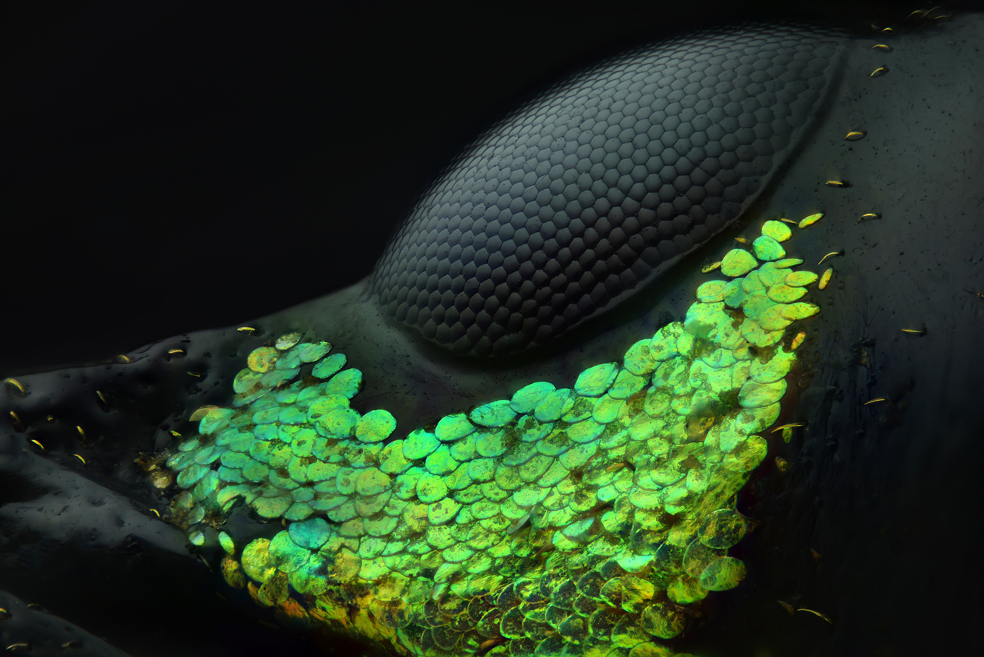
From Abu Dhabi, United Arab Emirates, photographer Yousef Al Habshi secured first place in the 2018 Nikon Small World photography contest using reflected light at a magnification of 20x to record this image of the eye of a Metapocyrtus subquadrulifer beetle. [Read more about the 2018 Nikon Small World winners]
Spore containers
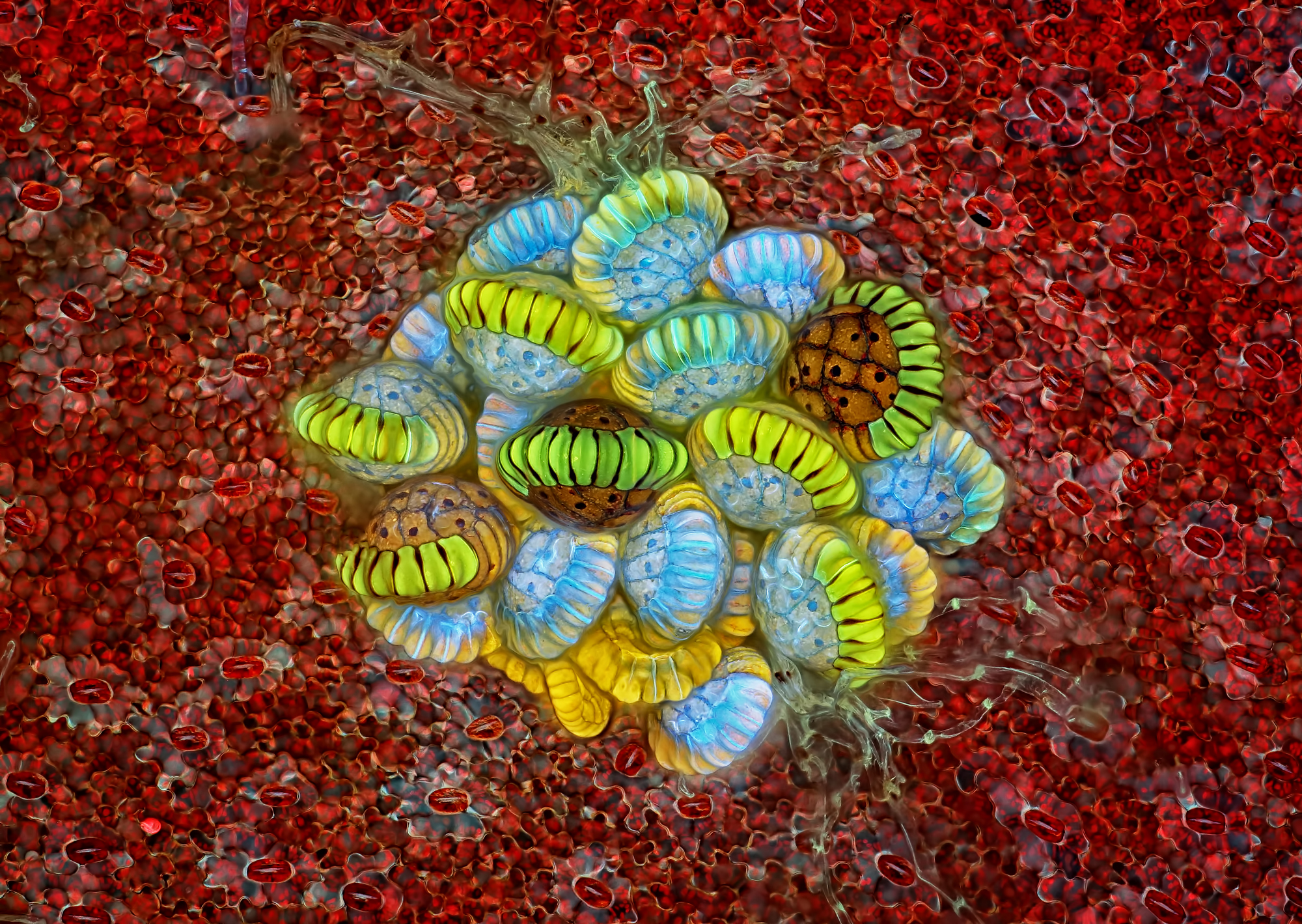
Rogelio Moreno from Panama City, Panama, used the photography technique of autofluorescence at 10x magnification to garner second place in the photography contest. The image exhibits a fern sorus with structures that produce and contain spores.
Spittlebug bubbles
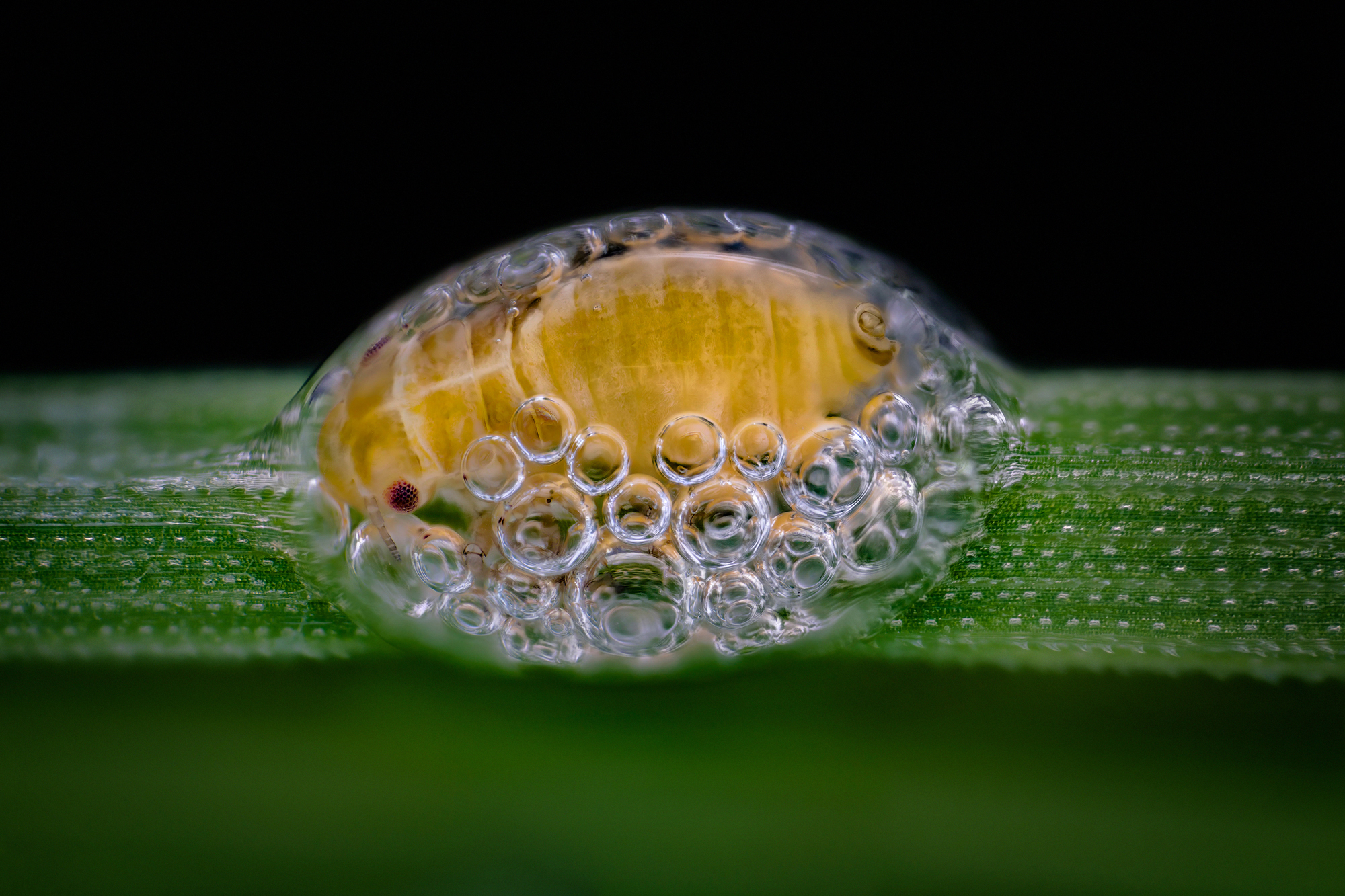
Photographer Saulius Gugis of Naperville, Illinois, took home third place in the 2018 Nikon Small World photography contest. The image of a spittlebug nymph in its bubble house utilized focus stacking at 5x magnification.
Peacock fronds
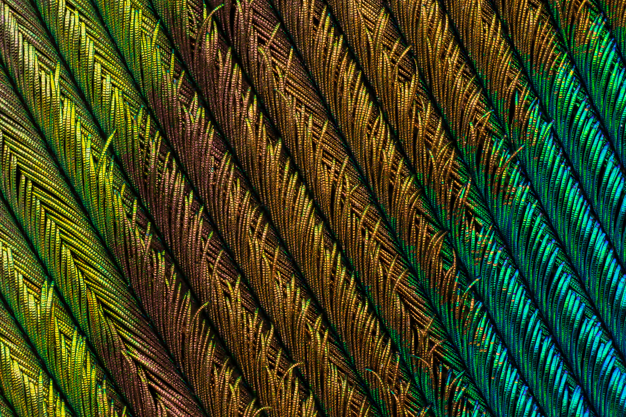
Claiming fourth place in the 2018 Nikon Small World photography contest, photographer Can Tunçer from Izmir, Turkey, captured this image of a peacock feather section. The image was created using focus stacking at a magnification of 5x.
Spider embryo
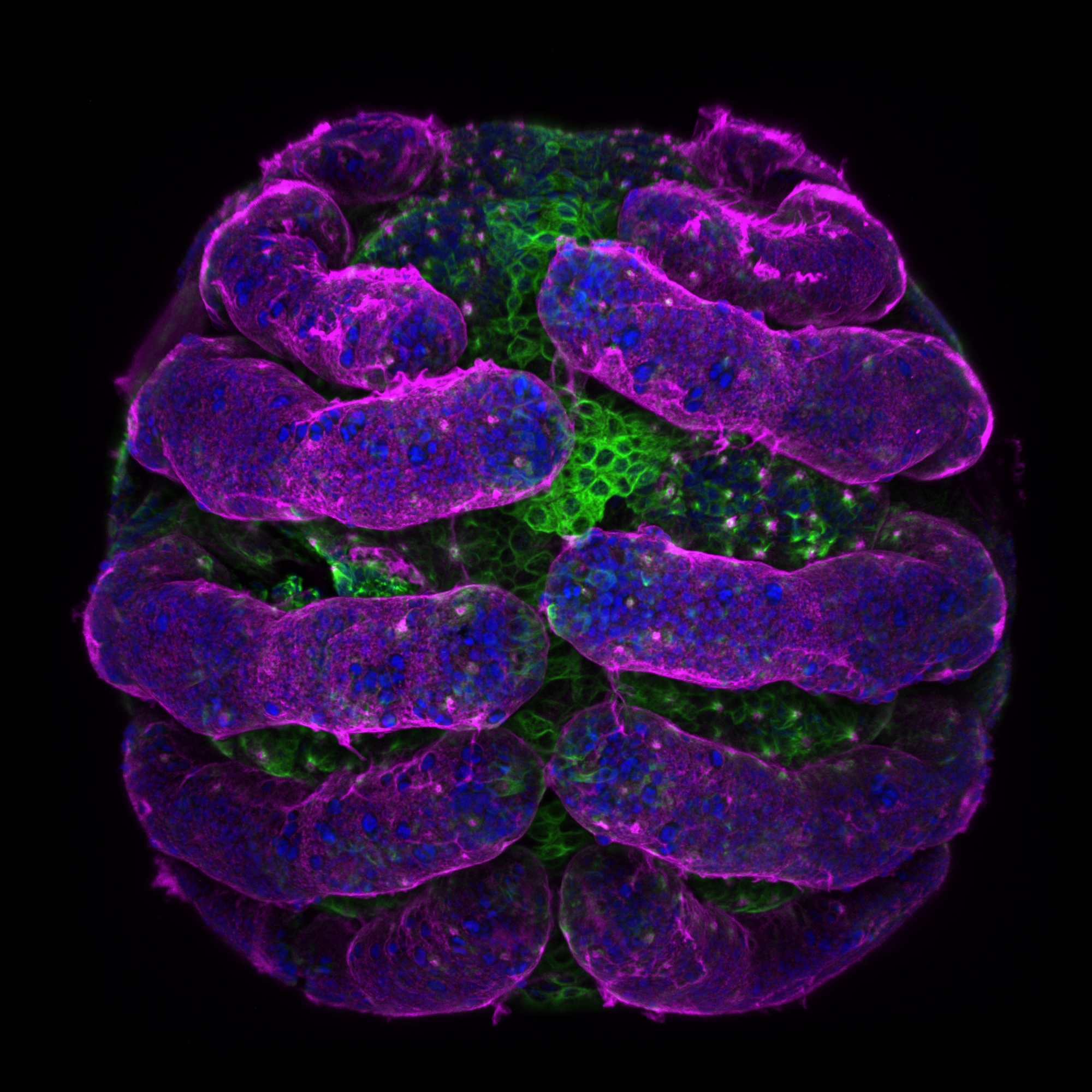
From Cambridge, Massachusetts, photographer Tessa Montague garnered fifth place using the confocal photography technique at a magnification of 20x to record this image of a Parasteatoda tepidariorum — a spider embryo — in the annual Nikon Small World photography competition. The image displays the embryo surface in pink, the nuclei in blue and the microtubules in green.
Retinal "explosion"
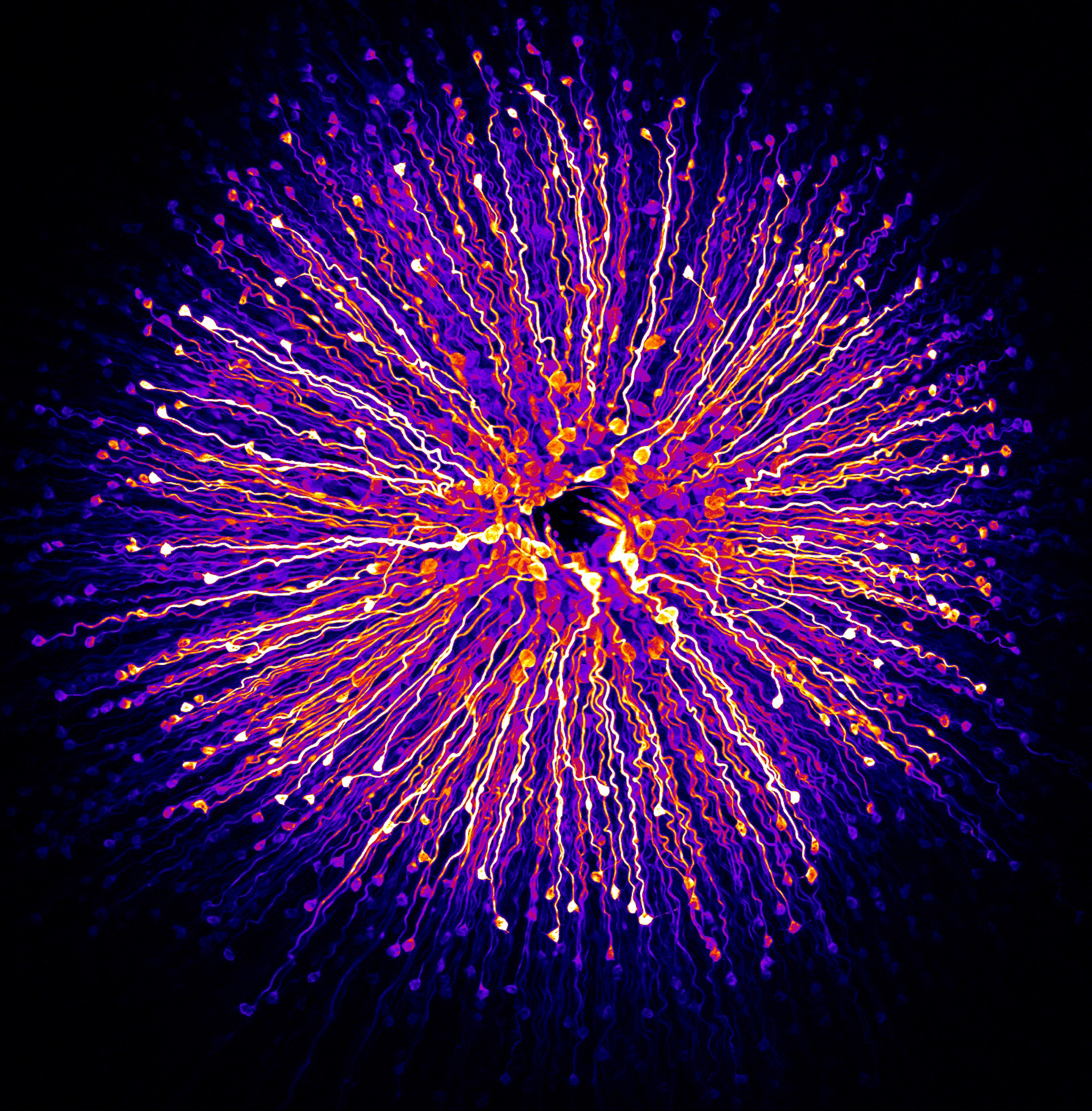
Hailing from the Vision Institute, Department of Therapeutics, a research center for eye diseases in Paris, France, Hanen Khabou used fluorescence and 40x magnification to take home sixth place in the photography contest. The image displays primate foveola — the central region of the retina.
Human teardrop
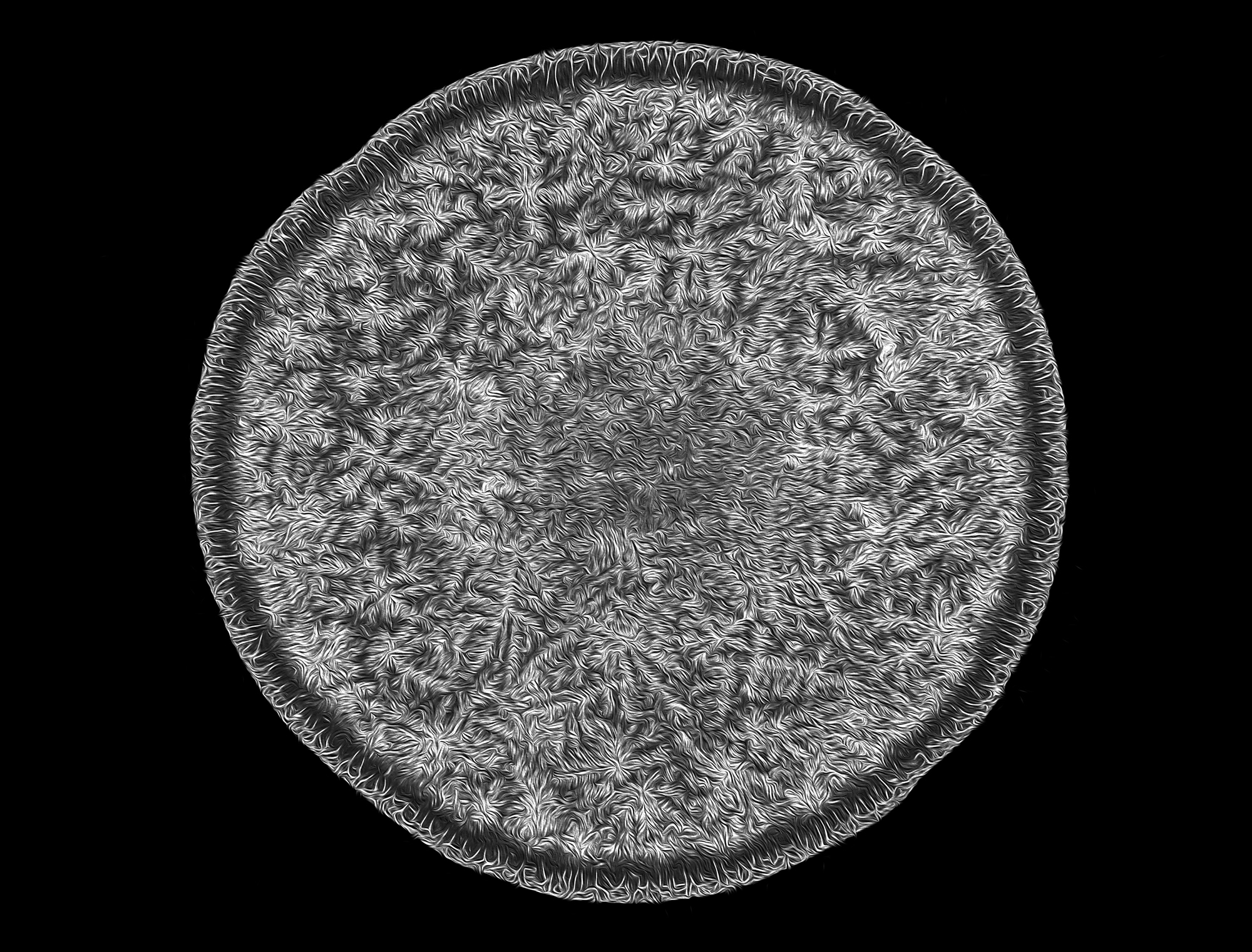
Photographer Norm Barker at the Johns Hopkins School of Medicine in Baltimore, Maryland, captured seventh place in the 2018 Nikon Small World photography contest. This image of a human teardrop utilized the darkfield technique — creating contrast in unstained specimens — at 5x magnification.
Sign up for the Live Science daily newsletter now
Get the world’s most fascinating discoveries delivered straight to your inbox.
Scaly face
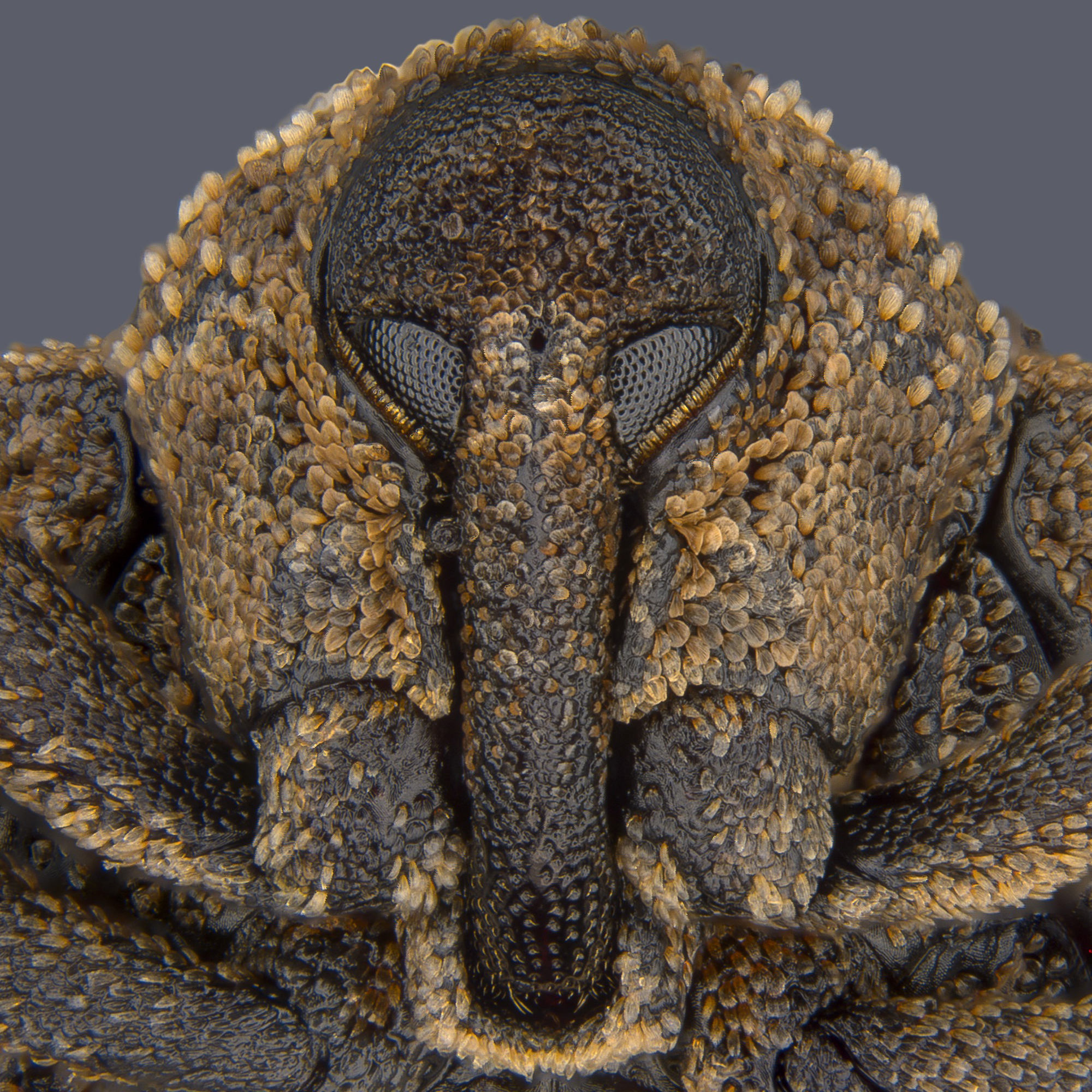
Taking eighth place in the 2018 Nikon Small World photography contest was Pia Scanlon of the Government of Western Australia, Department of Primary Industries and Regional Development. Scanlon submitted this portrait of a Sternochetus mangiferae, also known as a mango seed weevil. Using stereomicroscopy and image stacking to produce remarkable detail, Scanlon displayed the tiny insect at only 1x magnification.
Hologram circles
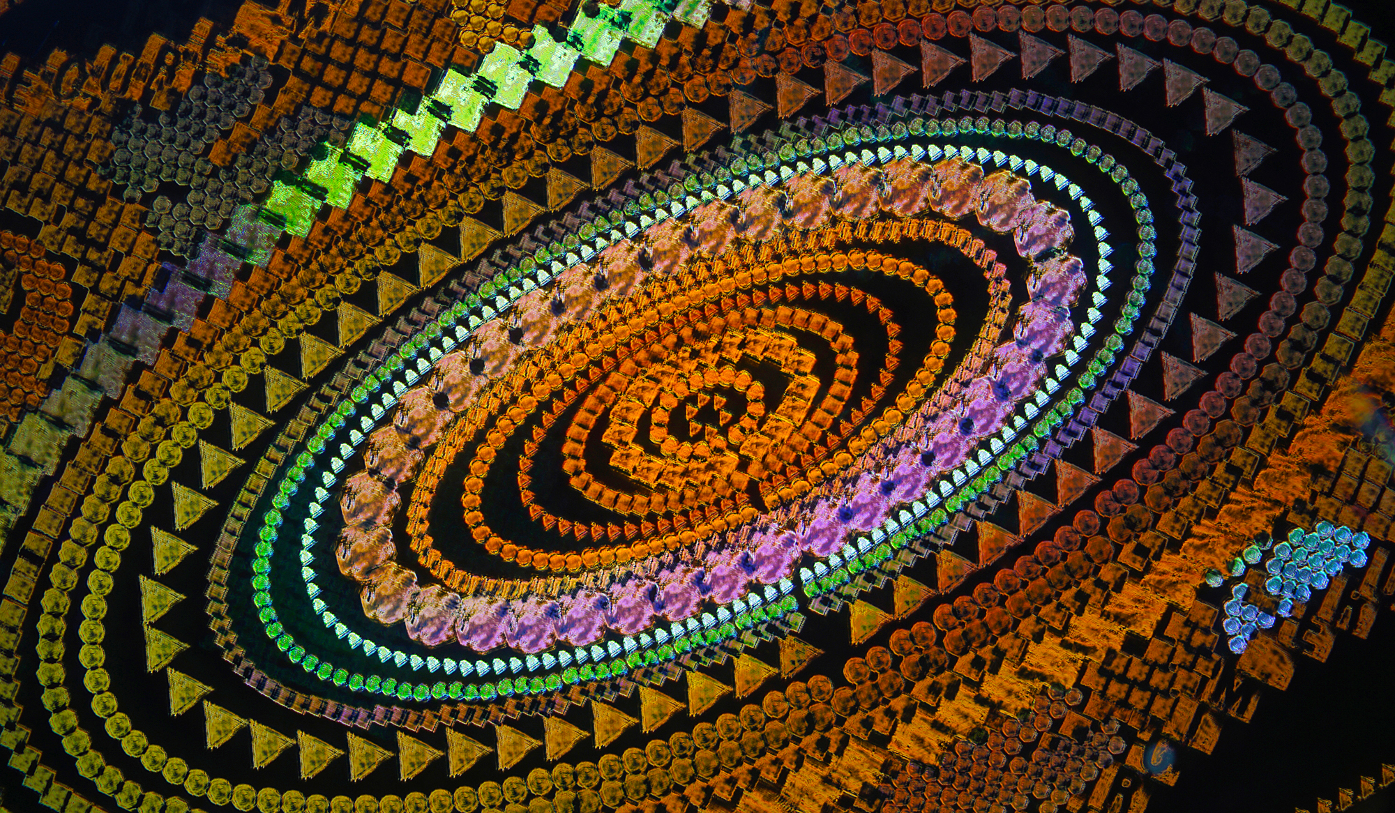
Haris Antonopoulos from Athens, Greece garnered 9th place using darkfield epi-illumination — reflected light microscopy. Antonopoulos used a magnification of 10x to record this image of a security hologram.
Pollen-studded
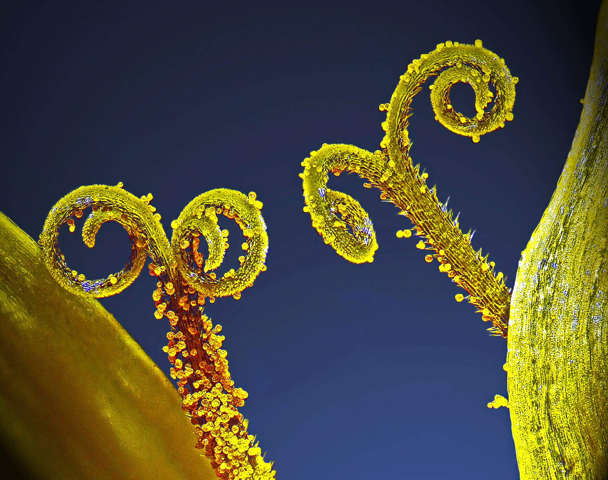
From the Department of Plant Protection at the University of Pannonia in Hungary, Csaba Pintér used the photography technique of focus stacking at 3x magnification to take home tenth place in the photography contest. The image presents plant stalks covered with pollen grains.
Cell division
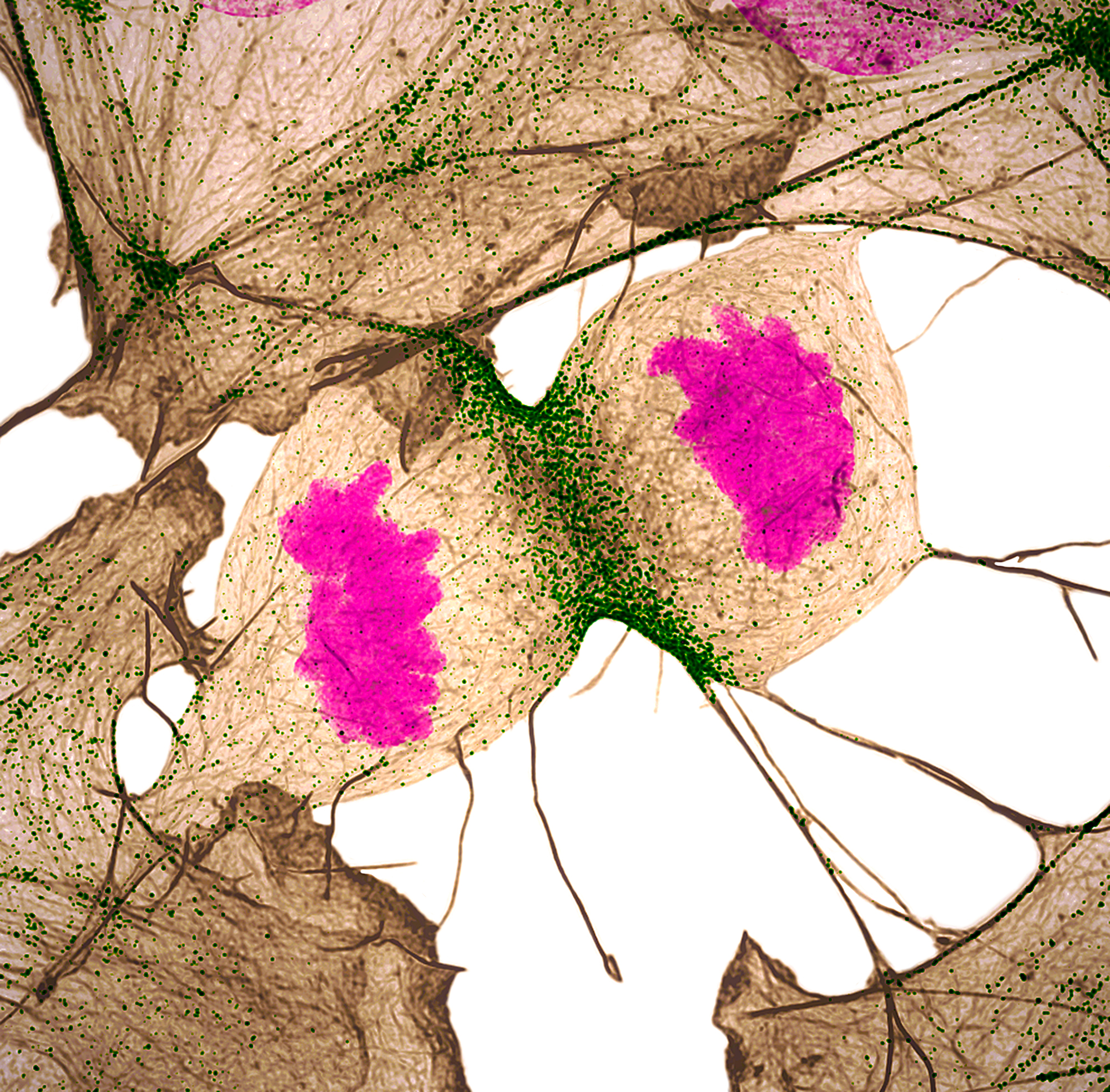
Photographers Nilay Taneja and Dylan Burnette with the Department of Cell and Developmental Biology at Vanderbilt University in Nashville, Tennessee, captured eleventh place in the 2018 Nikon Small World photography contest. Their image of a human fibroblast — a connective tissue cell —undergoing cell division utilized structured illumination microscopy at 60x magnification. In the image the actin protein displays gray, myosin II proteins show up as green, and DNA glows magenta.

