Strange purple lump on infant's head is rare case of protruding brain tissue, case report says
The cause of the boy's lump is unknown, although doctors suspect it could be an unusual symptom of a neural tube defect.

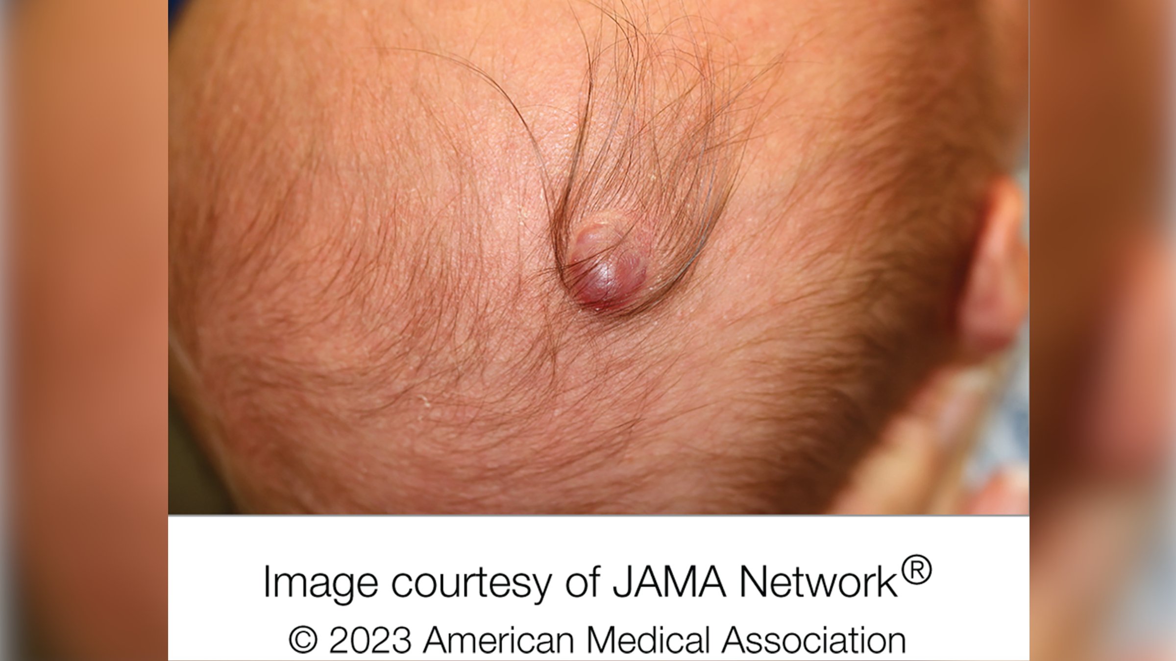
A boy who was born with a purple lump on his head had a rare skin defect and a bit of brain tissue spilling out of his skull, a new case report says.
The lump, which was on the right top of the boy's head, measured around 0.5 square inches (3.2 square centimeters) in area and had a ring of long, dark hairs surrounding it. This ring of hair, known as the "hair collar sign," can point to a type of neural tube defect, which happens when the neural tube — the structure from which the brain and spinal cord originate — doesn't close properly as a fetus develops in the womb. Such defects can cause protrusions of brain tissue to form lumps on the scalp.
Doctors involved in the case, published Sept. 20 in the journal JAMA Dermatology, diagnosed the child with a rare condition called aplasia cutis congenita (ACC). Believed to affect just 1 to 3 in 10,000 children, ACC causes babies to be born without skin on part of the body, most typically the scalp; structures under the skin, such as connective tissue or bone, can sometimes be missing too.
"Most cases typically occur on the scalp, appear to be an ulcer or wound and may be surrounded by extra long hair," Dr. Jenna Borok, lead author of the case report and pediatric dermatologist at the Ann & Robert H. Lurie Children's Hospital of Chicago, told Live Science in an email.
Related: Woman's 'extra breast' under her armpit developed a wart-like tumor in unusual case
Doctors don't know what causes ACC, but several factors may contribute to its development, including infection or physical trauma in the womb, exposure to drugs that can cause birth defects, and neural tube defects. Mutations in a gene called BMS1, which is involved in building proteins in cells, have also been linked to the condition.
The boy in the report had a specific type of ACC called bullous ACC, which typically starts out as a cyst-like lump that can be filled with fluid and then shrinks into a smaller plaque that is covered by a thin membrane.
Sign up for the Live Science daily newsletter now
Get the world’s most fascinating discoveries delivered straight to your inbox.
At 2-weeks-old, the boy had been diagnosed with bullous ACC with hair collar sign, in reference to the ring of hair around the lump; however, initial scans of his head didn't suggest any brain tissue was in the lump.
But when he reached 6 years of age, doctors surgically removed the lump and found that the extracted tissue showed signs of encephalocele, a neural tube defect where brain tissue projects through a hole in the skull and forms a sac-like structure on the head. So his diagnosis was tweaked to bullous ACC with encephalocele.
The boy's case and six previously reported cases likely support the idea that bullous ACC is an unusual manifestation of a neural tube defect, the case report authors wrote. The authors didn't detail if the boy had any intellectual or physical impairments as a result of the condition.
In terms of treatment, some ACC cases involve surgically altering the appearance of the resulting scar, and sometimes skin or bone grafting is needed to repair larger defects, the authors wrote in the report.
This article is for informational purposes only and is not meant to offer medical advice.

Emily is a health news writer based in London, United Kingdom. She holds a bachelor's degree in biology from Durham University and a master's degree in clinical and therapeutic neuroscience from Oxford University. She has worked in science communication, medical writing and as a local news reporter while undertaking NCTJ journalism training with News Associates. In 2018, she was named one of MHP Communications' 30 journalists to watch under 30. (emily.cooke@futurenet.com)
Flu: Facts about seasonal influenza and bird flu
What is hantavirus? The rare but deadly respiratory illness spread by rodents


