Creepy images show airway cells teeming with SARS-CoV-2
The images show a close-up view of the novel coronavirus infecting cells that line the human airway.
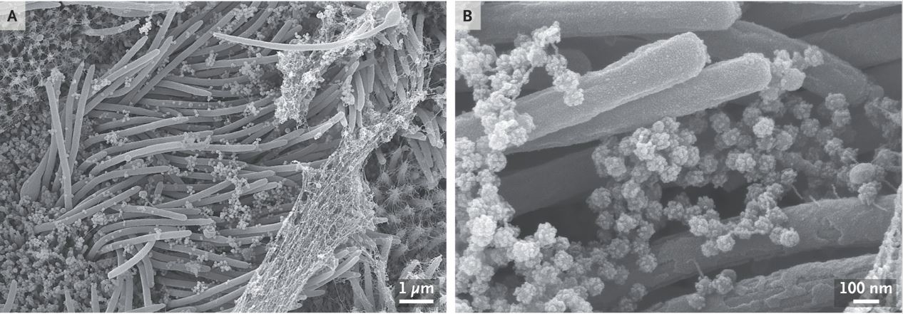
Striking new images show a close-up view of the novel coronavirus as it invades cells that line the human airway. The images provide a glimpse of the staggering number of virus particles that are produced during an infection — indeed, infected cells can churn out virus particles by the thousands.
Camille Ehre, an assistant professor at University of North Carolina School of Medicine's Marsico Lung Institute, captured the images with a scanning electron microscope, a device that uses a focused beam of electrons to produce images. To obtain the images, published Wednesday (Sept. 2) in The New England Journal of Medicine, researchers first infected human airway cells with SARS-CoV-2 — the coronavirus that causes COVID-19 — in lab dishes, and then examined the cells after four days.
The noodle-like projections in the images are cilia, or hair-like structures on the surface of some airway cells. The tips of the cilia are attached to strands of net-like mucus, which is naturally present in the airway.
The SARS-CoV-2 virus particles look like teensy balls. At a high magnification, you can make out the spiky structures on the surfaces of the virus particles. Those crown-like spikes give the family of "coronaviruses" its name. ("Corona" means "crown" in Latin.) The virus uses those spikes to invade human cells.
The image also shows a high density of virus particles, or virions. An analysis of the concentration of virus in the sample suggests a "high number of virions produced and released per [airway] cell," Ehre wrote.
Originally published on Live Science.
Sign up for the Live Science daily newsletter now
Get the world’s most fascinating discoveries delivered straight to your inbox.

Rachael is a Live Science contributor, and was a former channel editor and senior writer for Live Science between 2010 and 2022. She has a master's degree in journalism from New York University's Science, Health and Environmental Reporting Program. She also holds a B.S. in molecular biology and an M.S. in biology from the University of California, San Diego. Her work has appeared in Scienceline, The Washington Post and Scientific American.


